 Movie
Movie Controller

[English] 日本語
 Yorodumi
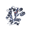
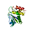
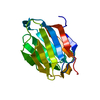
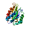
- PDB-3f8k: Crystal structure of protein acetyltransferase (PAT) from Sulfolo... -
+ Open data

- Basic information
| Entry | Database: PDB / ID: 3f8k | ||||||
|---|---|---|---|---|---|---|---|
| Title | Crystal structure of protein acetyltransferase (PAT) from Sulfolobus solfataricus | ||||||
 Components Components | Protein acetyltransferase | ||||||
 Keywords Keywords |  TRANSFERASE / GCN5-related N-acetyltransferase TRANSFERASE / GCN5-related N-acetyltransferase | ||||||
| Function / homology |  Function and homology information Function and homology information | ||||||
| Biological species |   Sulfolobus solfataricus P2 (archaea) Sulfolobus solfataricus P2 (archaea) | ||||||
| Method | X-RAY DIFFRACTION / SYNCHROTRON / MAD / Resolution: 1.84 Å | ||||||
 Authors Authors | Brent, M.M. | ||||||
 Citation Citation | Journal: J.Biol.Chem. / Year: 2009 Title: Structure and Biochemical Characterization of Protein Acetyltransferase from Sulfolobus solfataricus. Authors: Brent, M.M. / Iwata, A. / Carten, J. / Zhao, K. / Marmorstein, R. | ||||||
| History |
|
- Structure visualization
| Structure viewer | Molecule: MolmilJmol/JSmol |
|---|
- Downloads & links
-Download
| PDBx/mmCIF format | 3f8k.cif.gz | 42.1 KB | Display | PDBx/mmCIF format |
|---|---|---|---|---|
| PDB format | pdb3f8k.ent.gz | 28.9 KB | Display | PDB format |
| PDBx/mmJSON format | 3f8k.json.gz | Tree view | PDBx/mmJSON format | |
| Others |  Other downloads Other downloads |
-Validation report
| Arichive directory | https://data.pdbj.org/pub/pdb/validation_reports/f8/3f8kftp://data.pdbj.org/pub/pdb/validation_reports/f8/3f8k | HTTPS FTP |
|---|
-Related structure data
| Similar structure data |
|---|


-Links
 PDBj
PDBj


- Assembly
| Deposited unit | 
| ||||||||
|---|---|---|---|---|---|---|---|---|---|
| 1 |
| ||||||||
| Unit cell |
|
-Components
| #1: Protein | Mass: 18818.879 Da / Num. of mol.: 1 Source method: isolated from a genetically manipulated source Source: (gene. exp.) Sulfolobus solfataricus P2 (archaea) / Strain: DSM 1617 / JCM 11322 / P2 / Gene: SSO2813 / Plasmid: pET28a / Production host:  Escherichia coli (E. coli) / Strain (production host): BL21(DE3) / References: UniProt: Q97V23 Escherichia coli (E. coli) / Strain (production host): BL21(DE3) / References: UniProt: Q97V23 |
|---|---|
| #2: Chemical | ChemComp-COA / Coenzyme A  Mass: 767.534 Da / Num. of mol.: 1 / Source method: obtained synthetically / Formula: C21H36N7O16P3S Mass: 767.534 Da / Num. of mol.: 1 / Source method: obtained synthetically / Formula: C21H36N7O16P3S |
| #3: Water | ChemComp-HOH / Water Mass: 18.015 Da / Num. of mol.: 73 / Source method: isolated from a natural source / Formula: H2O Mass: 18.015 Da / Num. of mol.: 73 / Source method: isolated from a natural source / Formula: H2O |
-Experimental details
-Experiment
| Experiment | Method: X-RAY DIFFRACTION / Number of used crystals: 1 |
|---|
- Sample preparation
| Crystal | Density Matthews: 1.91 Å3/Da / Density % sol: 35.44 % Description: The structure factor file contains Friedel pairs |
|---|---|
| Crystal grow | Temperature: 300 K / Method: vapor diffusion, hanging drop / pH: 6.5 Details: 12% PEG 20000, 0.05M MES pH 6.5, VAPOR DIFFUSION, HANGING DROP, temperature 300K |
-Data collection
| Diffraction | Mean temperature: 100 K | ||||||||||||
|---|---|---|---|---|---|---|---|---|---|---|---|---|---|
| Diffraction source | Source: SYNCHROTRON / Site: APS  / Beamline: 23-ID-D / Wavelength: 0.97932, 0.97945, 0.94932 / Beamline: 23-ID-D / Wavelength: 0.97932, 0.97945, 0.94932 | ||||||||||||
| Detector | Type: MARMOSAIC 325 mm CCD / Detector: CCD / Date: Jul 3, 2006 / Details: Adjustable focus K-B pair Si plus Pt, Rh coatings | ||||||||||||
| Radiation | Monochromator: Double crystal cryo-cooled Si(111) / Protocol: MAD / Monochromatic (M) / Laue (L): M / Scattering type: x-ray | ||||||||||||
| Radiation wavelength |
| ||||||||||||
| Reflection | Resolution: 1.84→30 Å / Num. all: 23855 / Num. obs: 23521 / % possible obs: 98.6 % / Redundancy: 5.9 % / Rmerge(I) obs: 0.068 / Net I/σ(I): 23.1 | ||||||||||||
| Reflection shell | Resolution: 1.84→1.92 Å / Redundancy: 3.8 % / Rmerge(I) obs: 0.423 / Mean I/σ(I) obs: 2.5 / % possible all: 89.2 |
- Processing
| Software |
| ||||||||||||||||||||||||||||
|---|---|---|---|---|---|---|---|---|---|---|---|---|---|---|---|---|---|---|---|---|---|---|---|---|---|---|---|---|---|
| Refinement | Method to determine structure: MAD / Resolution: 1.84→30 Å / Details: The Friedel pairs were used in phasing
| ||||||||||||||||||||||||||||
| Solvent computation | Bsol: 68.0955 Å2 / ksol: 0.415774 e/Å3 | ||||||||||||||||||||||||||||
| Displacement parameters |
| ||||||||||||||||||||||||||||
| Refinement step | Cycle: LAST / Resolution: 1.84→30 Å
| ||||||||||||||||||||||||||||
| Refine LS restraints |
|
