 Movie
Movie Controller


+ Open data

- Basic information
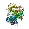
| Entry | Database: PDB / ID: 3f88 | ||||||
|---|---|---|---|---|---|---|---|
| Title | glycogen synthase Kinase 3beta inhibitor complex | ||||||
 Components Components | Glycogen synthase kinase-3 beta | ||||||
 Keywords Keywords | TRANSFERASE / enzyme / protein kinase / inhibitor / Alternative splicing / ATP-binding / Kinase / Nucleotide-binding / Phosphoprotein / Serine/threonine-protein kinase / Wnt signaling pathway | ||||||
| Function / homology |  Function and homology information Function and homology informationnegative regulation of glycogen (starch) synthase activity / neuron projection organization / regulation of microtubule anchoring at centrosome / beta-catenin destruction complex disassembly / negative regulation of mesenchymal stem cell differentiation / negative regulation of type B pancreatic cell development / superior temporal gyrus development / positive regulation of protein localization to cilium / negative regulation of glycogen biosynthetic process / negative regulation of dopaminergic neuron differentiation ...negative regulation of glycogen (starch) synthase activity / neuron projection organization / regulation of microtubule anchoring at centrosome / beta-catenin destruction complex disassembly / negative regulation of mesenchymal stem cell differentiation / negative regulation of type B pancreatic cell development / superior temporal gyrus development / positive regulation of protein localization to cilium / negative regulation of glycogen biosynthetic process / negative regulation of dopaminergic neuron differentiation / maintenance of cell polarity / positive regulation of protein localization to centrosome / : / positive regulation of mitochondrial outer membrane permeabilization involved in apoptotic signaling pathway / positive regulation of cilium assembly / negative regulation of protein acetylation / APC truncation mutants have impaired AXIN binding / AXIN missense mutants destabilize the destruction complex / Truncations of AMER1 destabilize the destruction complex / beta-catenin destruction complex / tau-protein kinase / CRMPs in Sema3A signaling / regulation of microtubule-based process / heart valve development / regulation of protein export from nucleus / Beta-catenin phosphorylation cascade / Signaling by GSK3beta mutants / CTNNB1 S33 mutants aren't phosphorylated / CTNNB1 S37 mutants aren't phosphorylated / CTNNB1 S45 mutants aren't phosphorylated / CTNNB1 T41 mutants aren't phosphorylated / Maturation of nucleoprotein / cellular response to interleukin-3 / Wnt signalosome / negative regulation of protein localization to nucleus / regulation of long-term synaptic potentiation / Disassembly of the destruction complex and recruitment of AXIN to the membrane / Maturation of nucleoprotein / AKT phosphorylates targets in the cytosol / negative regulation of calcineurin-NFAT signaling cascade / positive regulation of cell-matrix adhesion / regulation of axon extension / dopamine receptor signaling pathway / negative regulation of phosphoprotein phosphatase activity / regulation of dendrite morphogenesis / regulation of axonogenesis / establishment of cell polarity / tau-protein kinase activity / glycogen metabolic process / ER overload response / regulation of neuron projection development / Constitutive Signaling by AKT1 E17K in Cancer / protein kinase A catalytic subunit binding / dynactin binding / NF-kappaB binding / canonical Wnt signaling pathway / Regulation of HSF1-mediated heat shock response / epithelial to mesenchymal transition / negative regulation of osteoblast differentiation / negative regulation of protein-containing complex assembly / positive regulation of autophagy / regulation of microtubule cytoskeleton organization / regulation of cellular response to heat / cellular response to retinoic acid / negative regulation of extrinsic apoptotic signaling pathway via death domain receptors / extrinsic apoptotic signaling pathway / extrinsic apoptotic signaling pathway in absence of ligand / excitatory postsynaptic potential / presynaptic modulation of chemical synaptic transmission / positive regulation of protein export from nucleus / positive regulation of protein ubiquitination / Ubiquitin-dependent degradation of Cyclin D / hippocampus development / positive regulation of cell differentiation / positive regulation of protein-containing complex assembly / GSK3B and BTRC:CUL1-mediated-degradation of NFE2L2 / peptidyl-threonine phosphorylation / Degradation of GLI2 by the proteasome / GLI3 is processed to GLI3R by the proteasome / Degradation of beta-catenin by the destruction complex / tau protein binding / negative regulation of canonical Wnt signaling pathway / regulation of circadian rhythm / B-WICH complex positively regulates rRNA expression / beta-catenin binding / positive regulation of GTPase activity / circadian rhythm / cellular response to amyloid-beta / positive regulation of protein catabolic process / Regulation of RUNX2 expression and activity / neuron projection development / positive regulation of proteasomal ubiquitin-dependent protein catabolic process / p53 binding / presynapse / positive regulation of protein binding / insulin receptor signaling pathway / kinase activity / postsynapse / proteasome-mediated ubiquitin-dependent protein catabolic process / peptidyl-serine phosphorylationSimilarity search - Function | ||||||
| Biological species |  Homo sapiens (human) Homo sapiens (human) | ||||||
| Method | X-RAY DIFFRACTION / SYNCHROTRON / Resolution: 2.6 Å | ||||||
 Authors Authors | Mol, C.D. / Dougan, D.R. | ||||||
 Citation Citation | Journal: Bioorg.Med.Chem. / Year: 2009 Title: Design, synthesis and structure-activity relationships of 1,3,4-oxadiazole derivatives as novel inhibitors of glycogen synthase kinase-3beta. Authors: Saitoh, M. / Kunitomo, J. / Kimura, E. / Hayase, Y. / Kobayashi, H. / Uchiyama, N. / Kawamoto, T. / Tanaka, T. / Mol, C.D. / Dougan, D.R. / Textor, G.S. / Snell, G.P. / Itoh, F. | ||||||
| History |
|
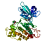
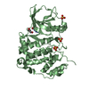
- Structure visualization
| Structure viewer | Molecule: MolmilJmol/JSmol |
|---|
- Downloads & links
-Download
| PDBx/mmCIF format | 3f88.cif.gz | 152.4 KB | Display | PDBx/mmCIF format |
|---|---|---|---|---|
| PDB format | pdb3f88.ent.gz | 120.1 KB | Display | PDB format |
| PDBx/mmJSON format | 3f88.json.gz | Tree view | PDBx/mmJSON format | |
| Others |  Other downloads Other downloads |
-Validation report
| Arichive directory | https://data.pdbj.org/pub/pdb/validation_reports/f8/3f88ftp://data.pdbj.org/pub/pdb/validation_reports/f8/3f88 | HTTPS FTP |
|---|
-Related structure data


-Links
 PDBj
PDBj


- Assembly
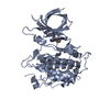
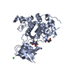
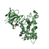
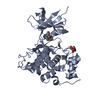
| Deposited unit | 
| ||||||||
|---|---|---|---|---|---|---|---|---|---|
| 1 | 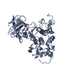
| ||||||||
| 2 | 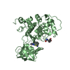
| ||||||||
| Unit cell |
|
-Components
| #1: Protein | / GSK-3 beta Mass: 39739.633 Da / Num. of mol.: 2 / Fragment: UNP residues 35-383, Protein kinase domain Source method: isolated from a genetically manipulated source Source: (gene. exp.) Homo sapiens (human) / Gene: GSK3B / Production host:   Spodoptera frugiperda (fall armyworm) / Strain (production host): Hi5 / References: UniProt: P49841, tau-protein kinase Spodoptera frugiperda (fall armyworm) / Strain (production host): Hi5 / References: UniProt: P49841, tau-protein kinase#2: Chemical |   Mass: 324.357 Da / Num. of mol.: 2 / Source method: obtained synthetically / Formula: C16H12N4O2S Mass: 324.357 Da / Num. of mol.: 2 / Source method: obtained synthetically / Formula: C16H12N4O2S#3: Chemical |   Mass: 117.148 Da / Num. of mol.: 2 / Source method: obtained synthetically / Formula: C8H7N Mass: 117.148 Da / Num. of mol.: 2 / Source method: obtained synthetically / Formula: C8H7N#4: Water | ChemComp-HOH / | Water Mass: 18.015 Da / Num. of mol.: 145 / Source method: isolated from a natural source / Formula: H2O Mass: 18.015 Da / Num. of mol.: 145 / Source method: isolated from a natural source / Formula: H2O |
|---|
-Experimental details
-Experiment
| Experiment | Method: X-RAY DIFFRACTION / Number of used crystals: 1 |
|---|
- Sample preparation
| Crystal | Density Matthews: 3.1 Å3/Da / Density % sol: 60.27 % |
|---|---|
| Crystal grow | Temperature: 277 K / Method: vapor diffusion, sitting drop / pH: 7.5 Details: 10% PEG 3350, 0.2M proline, 0.1M HEPES, pH 7.5, VAPOR DIFFUSION, SITTING DROP, temperature 277K |
-Data collection
| Diffraction | Mean temperature: 100 K |
|---|---|
| Diffraction source | Source: SYNCHROTRON / Site: ALS  / Beamline: 5.0.3 / Wavelength: 1 Å / Beamline: 5.0.3 / Wavelength: 1 Å |
| Detector | Type: ADSC QUANTUM 315 / Detector: CCD / Date: Jul 21, 2006 / Details: mirrors |
| Radiation | Monochromator: SAGITALLY FOCUSED Si(111) / Protocol: SINGLE WAVELENGTH / Monochromatic (M) / Laue (L): M / Scattering type: x-ray |
| Radiation wavelength | Wavelength: 1 Å / Relative weight: 1 |
| Reflection | Resolution: 2.6→20 Å / Num. obs: 29408 / % possible obs: 98.5 % / Redundancy: 3.7 % / Rmerge(I) obs: 0.044 / Χ2: 1.193 / Net I/σ(I): 32.857 |
| Reflection shell | Resolution: 2.6→2.69 Å / Redundancy: 3.4 % / Rmerge(I) obs: 0.447 / Num. unique all: 2804 / Χ2: 0.769 / % possible all: 94.2 |
- Processing
| Software |
| ||||||||||||||||||||||||||||||||||||||||||||||||||||||||||||||||||||||||||||||||||||||||||
|---|---|---|---|---|---|---|---|---|---|---|---|---|---|---|---|---|---|---|---|---|---|---|---|---|---|---|---|---|---|---|---|---|---|---|---|---|---|---|---|---|---|---|---|---|---|---|---|---|---|---|---|---|---|---|---|---|---|---|---|---|---|---|---|---|---|---|---|---|---|---|---|---|---|---|---|---|---|---|---|---|---|---|---|---|---|---|---|---|---|---|---|
| Refinement | Resolution: 2.6→20 Å / Cor.coef. Fo:Fc: 0.96 / Cor.coef. Fo:Fc free: 0.92 / Occupancy max: 1 / Occupancy min: 0.35 / SU B: 32.423 / SU ML: 0.33 / Cross valid method: THROUGHOUT / σ(F): 0 / ESU R: 0.561 / ESU R Free: 0.338 / Stereochemistry target values: MAXIMUM LIKELIHOOD
| ||||||||||||||||||||||||||||||||||||||||||||||||||||||||||||||||||||||||||||||||||||||||||
| Solvent computation | Ion probe radii: 0.8 Å / Shrinkage radii: 0.8 Å / VDW probe radii: 1.2 Å / Solvent model: BABINET MODEL WITH MASK | ||||||||||||||||||||||||||||||||||||||||||||||||||||||||||||||||||||||||||||||||||||||||||
| Displacement parameters | Biso max: 158.25 Å2 / Biso mean: 75.643 Å2 / Biso min: 8 Å2
| ||||||||||||||||||||||||||||||||||||||||||||||||||||||||||||||||||||||||||||||||||||||||||
| Refine analyze | Luzzati coordinate error obs: 0 Å | ||||||||||||||||||||||||||||||||||||||||||||||||||||||||||||||||||||||||||||||||||||||||||
| Refinement step | Cycle: LAST / Resolution: 2.6→20 Å
| ||||||||||||||||||||||||||||||||||||||||||||||||||||||||||||||||||||||||||||||||||||||||||
| Refine LS restraints |
| ||||||||||||||||||||||||||||||||||||||||||||||||||||||||||||||||||||||||||||||||||||||||||
| LS refinement shell | Resolution: 2.6→2.666 Å / Total num. of bins used: 20
|
