 Movie
Movie Controller

[English] 日本語
 Yorodumi
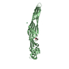
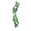
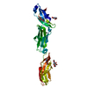
- PDB-3f6n: Crystal structure of the virion-associated protein P3 from Caulim... -
+ Open data

- Basic information
| Entry | Database: PDB / ID: 3f6n | ||||||
|---|---|---|---|---|---|---|---|
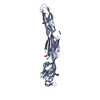
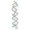
| Title | Crystal structure of the virion-associated protein P3 from Caulimovirus | ||||||
 Components Components | Virion-associated protein | ||||||
 Keywords Keywords |  VIRAL PROTEIN / DNA-BINDING PROTEIN / COILED-COIL / TETRAMER / DNA-binding / Virion VIRAL PROTEIN / DNA-BINDING PROTEIN / COILED-COIL / TETRAMER / DNA-binding / Virion | ||||||
| Function / homology |  Function and homology information Function and homology information | ||||||
| Biological species | Cauliflower mosaic virus | ||||||
| Method | X-RAY DIFFRACTION / SYNCHROTRON / MOLECULAR REPLACEMENT / molecular replacement / Resolution: 3.1 Å | ||||||
 Authors Authors | Hoh, F. / Uzest, M. / Blanc, S. / Dumas, C. | ||||||
 Citation Citation | Journal: J.Virol. / Year: 2010 Title: Structural insights into the molecular mechanisms of cauliflower mosaic virus transmission by its insect vector. Authors: Hoh, F. / Uzest, M. / Drucker, M. / Plisson-Chastang, C. / Bron, P. / Blanc, S. / Dumas, C. | ||||||
| History |
|
- Structure visualization
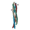
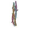
| Structure viewer | Molecule: MolmilJmol/JSmol |
|---|
- Downloads & links
-Download
| PDBx/mmCIF format | 3f6n.cif.gz | 67.2 KB | Display | PDBx/mmCIF format |
|---|---|---|---|---|
| PDB format | pdb3f6n.ent.gz | 49.5 KB | Display | PDB format |
| PDBx/mmJSON format | 3f6n.json.gz | Tree view | PDBx/mmJSON format | |
| Others |  Other downloads Other downloads |
-Validation report
| Arichive directory | https://data.pdbj.org/pub/pdb/validation_reports/f6/3f6nftp://data.pdbj.org/pub/pdb/validation_reports/f6/3f6n | HTTPS FTP |
|---|
-Related structure data
| Related structure data |  3k4tC  1t6fS S: Starting model for refinement C: citing same article ( |
|---|---|
| Similar structure data |


-Links
 PDBj
PDBj- Assembly
| Deposited unit | 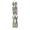
| |||||||||||||||||||||||||||||||||||||||||||||||||||||||||||||||||||||||||||||||||||||||||||||||||||||||||||||||||||||||||||||||||||||||||||||||||||||||||||||||||||||||||||||||||||||||||||||||||||||||||||
|---|---|---|---|---|---|---|---|---|---|---|---|---|---|---|---|---|---|---|---|---|---|---|---|---|---|---|---|---|---|---|---|---|---|---|---|---|---|---|---|---|---|---|---|---|---|---|---|---|---|---|---|---|---|---|---|---|---|---|---|---|---|---|---|---|---|---|---|---|---|---|---|---|---|---|---|---|---|---|---|---|---|---|---|---|---|---|---|---|---|---|---|---|---|---|---|---|---|---|---|---|---|---|---|---|---|---|---|---|---|---|---|---|---|---|---|---|---|---|---|---|---|---|---|---|---|---|---|---|---|---|---|---|---|---|---|---|---|---|---|---|---|---|---|---|---|---|---|---|---|---|---|---|---|---|---|---|---|---|---|---|---|---|---|---|---|---|---|---|---|---|---|---|---|---|---|---|---|---|---|---|---|---|---|---|---|---|---|---|---|---|---|---|---|---|---|---|---|---|---|---|---|---|---|---|
| 1 |
| |||||||||||||||||||||||||||||||||||||||||||||||||||||||||||||||||||||||||||||||||||||||||||||||||||||||||||||||||||||||||||||||||||||||||||||||||||||||||||||||||||||||||||||||||||||||||||||||||||||||||||
| Unit cell |
| |||||||||||||||||||||||||||||||||||||||||||||||||||||||||||||||||||||||||||||||||||||||||||||||||||||||||||||||||||||||||||||||||||||||||||||||||||||||||||||||||||||||||||||||||||||||||||||||||||||||||||
| Noncrystallographic symmetry (NCS) | NCS domain:
NCS domain segments: Component-ID: 1
|