 Movie
Movie Controller

[English] 日本語
 Yorodumi
- PDB-3eyc: New crystal structure of human tear lipocalin in complex with 1,4... -
+ Open data

- Basic information
| Entry | Database: PDB / ID: 3eyc | ||||||
|---|---|---|---|---|---|---|---|
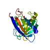
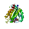
| Title | New crystal structure of human tear lipocalin in complex with 1,4-butanediol in space group P21 | ||||||
 Components Components | Lipocalin-1 | ||||||
 Keywords Keywords | TRANSPORT PROTEIN / ligand-binding protein / beta-barrel / Secreted / Sensory transduction / Taste / Transport | ||||||
| Function / homology |  Function and homology information Function and homology informationTransport of fatty acids / response to stimulus / chloride ion binding / cysteine-type endopeptidase inhibitor activity / small molecule binding / sensory perception of taste / signaling receptor binding / proteolysis / extracellular space / zinc ion binding / extracellular regionSimilarity search - Function | ||||||
| Biological species |  Homo sapiens (human) Homo sapiens (human) | ||||||
| Method | X-RAY DIFFRACTION / MOLECULAR REPLACEMENT / Resolution: 2.6 Å | ||||||
 Authors Authors | Breustedt, D.A. / Keil, L. / Skerra, A. | ||||||
 Citation Citation | Journal: Acta Crystallogr.,Sect.D / Year: 2009 Title: A new crystal form of human tear lipocalin reveals high flexibility in the loop region and induced fit in the ligand cavity Authors: Breustedt, D.A. / Chatwell, L. / Skerra, A. | ||||||
| History |
|
- Structure visualization
| Structure viewer | Molecule: MolmilJmol/JSmol |
|---|
- Downloads & links
-Download
| PDBx/mmCIF format | 3eyc.cif.gz | 126.4 KB | Display | PDBx/mmCIF format |
|---|---|---|---|---|
| PDB format | pdb3eyc.ent.gz | 96.9 KB | Display | PDB format |
| PDBx/mmJSON format | 3eyc.json.gz | Tree view | PDBx/mmJSON format | |
| Others |  Other downloads Other downloads |
-Validation report
| Arichive directory | https://data.pdbj.org/pub/pdb/validation_reports/ey/3eycftp://data.pdbj.org/pub/pdb/validation_reports/ey/3eyc | HTTPS FTP |
|---|
-Related structure data
| Related structure data |  1xkiS S: Starting model for refinement |
|---|---|
| Similar structure data |


-Links
 PDBj
PDBj


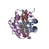
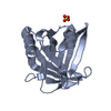
- Assembly
| Deposited unit | 
| ||||||||
|---|---|---|---|---|---|---|---|---|---|
| 1 | 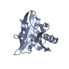
| ||||||||
| 2 | 
| ||||||||
| 3 | 
| ||||||||
| 4 | 
| ||||||||
| Unit cell |
|
-Components
| #1: Protein | / Von Ebner gland protein / VEG protein / Tear prealbumin / TP / Tear lipocalin / Tlc Mass: 17949.180 Da / Num. of mol.: 4 / Fragment: residues 5-166 / Mutation: C101S, D158A Source method: isolated from a genetically manipulated source Details: Tear / Source: (gene. exp.) Homo sapiens (human) / Gene: LCN1 / Plasmid: pTlc3 / Production host:  Escherichia coli (E. coli) / Strain (production host): JM83 / References: UniProt: P31025 Escherichia coli (E. coli) / Strain (production host): JM83 / References: UniProt: P31025#2: Chemical | ChemComp-BU1 / 1,4-Butanediol  Mass: 90.121 Da / Num. of mol.: 4 / Source method: obtained synthetically / Formula: C4H10O2 Mass: 90.121 Da / Num. of mol.: 4 / Source method: obtained synthetically / Formula: C4H10O2#3: Water | ChemComp-HOH / | Water Mass: 18.015 Da / Num. of mol.: 185 / Source method: isolated from a natural source / Formula: H2O Mass: 18.015 Da / Num. of mol.: 185 / Source method: isolated from a natural source / Formula: H2O |
|---|
-Experimental details
-Experiment
| Experiment | Method: X-RAY DIFFRACTION / Number of used crystals: 1 |
|---|
- Sample preparation
| Crystal | Density Matthews: 2.18 Å3/Da / Density % sol: 43.62 % / Mosaicity: 0.466 ° |
|---|---|
| Crystal grow | Temperature: 293 K / Method: vapor diffusion, hanging drop / pH: 8 Details: 27% PEG3350, 0.8% 1.4-Butanediol, 200mM NaCl, 100mM Tris/HCl, pH 8.0, VAPOR DIFFUSION, HANGING DROP, temperature 293K |
-Data collection
| Diffraction | Mean temperature: 100 K |
|---|---|
| Diffraction source | Source: ROTATING ANODE / Type: RIGAKU RU300 / Wavelength: 1.5418 Å |
| Detector | Type: MAR scanner 345 mm plate / Detector: IMAGE PLATE / Date: May 5, 2005 / Details: Osmics mirrors |
| Radiation | Monochromator: Graphite / Protocol: SINGLE WAVELENGTH / Monochromatic (M) / Laue (L): M / Scattering type: x-ray |
| Radiation wavelength | Wavelength: 1.5418 Å / Relative weight: 1 |
| Reflection | Resolution: 2.6→20 Å / Num. obs: 19625 / % possible obs: 99.6 % / Redundancy: 5.7 % / Rmerge(I) obs: 0.061 / Χ2: 1.037 / Net I/σ(I): 16 |
| Reflection shell | Resolution: 2.6→2.69 Å / % possible obs: 97 % / Redundancy: 4.8 % / Rmerge(I) obs: 0.236 / Num. unique obs: 1852 / Χ2: 0.991 |
- Processing
| Software |
| ||||||||||||||||||||||||||||||||||||||||||||||||||||||||||||||||||||||||||||||||||||||||||||||||||||||||||||||||||||||||||||||
|---|---|---|---|---|---|---|---|---|---|---|---|---|---|---|---|---|---|---|---|---|---|---|---|---|---|---|---|---|---|---|---|---|---|---|---|---|---|---|---|---|---|---|---|---|---|---|---|---|---|---|---|---|---|---|---|---|---|---|---|---|---|---|---|---|---|---|---|---|---|---|---|---|---|---|---|---|---|---|---|---|---|---|---|---|---|---|---|---|---|---|---|---|---|---|---|---|---|---|---|---|---|---|---|---|---|---|---|---|---|---|---|---|---|---|---|---|---|---|---|---|---|---|---|---|---|---|---|
| Refinement | Method to determine structure: MOLECULAR REPLACEMENT Starting model: 1xki, residues 14 to 24, 38 to 55, 64 to 104 and 110 to 150 Resolution: 2.6→20 Å / FOM work R set: 0.798 / Stereochemistry target values: Engh & Huber
| ||||||||||||||||||||||||||||||||||||||||||||||||||||||||||||||||||||||||||||||||||||||||||||||||||||||||||||||||||||||||||||||
| Displacement parameters | Biso mean: 43.683 Å2 | ||||||||||||||||||||||||||||||||||||||||||||||||||||||||||||||||||||||||||||||||||||||||||||||||||||||||||||||||||||||||||||||
| Refinement step | Cycle: LAST / Resolution: 2.6→20 Å
| ||||||||||||||||||||||||||||||||||||||||||||||||||||||||||||||||||||||||||||||||||||||||||||||||||||||||||||||||||||||||||||||
| Refine LS restraints |
| ||||||||||||||||||||||||||||||||||||||||||||||||||||||||||||||||||||||||||||||||||||||||||||||||||||||||||||||||||||||||||||||
| LS refinement shell | Refine-ID: X-RAY DIFFRACTION / Total num. of bins used: 20
|
