 Movie
Movie Controller


+ Open data

- Basic information
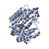
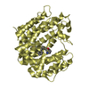
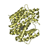
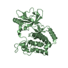
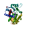
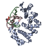
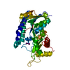
| Entry | Database: PDB / ID: 3eu7 | ||||||
|---|---|---|---|---|---|---|---|
| Title | Crystal Structure of a PALB2 / BRCA2 complex | ||||||
 Components Components |
| ||||||
 Keywords Keywords | Transcription/antitumor Protein / WD40 domain /  Beta Propeller / Protein-Peptide Complex / Fanconi anemia / Nucleus / Phosphoprotein / WD repeat / Disease mutation / DNA damage / DNA repair / Transcription-antitumor Protein COMPLEX Beta Propeller / Protein-Peptide Complex / Fanconi anemia / Nucleus / Phosphoprotein / WD repeat / Disease mutation / DNA damage / DNA repair / Transcription-antitumor Protein COMPLEX | ||||||
| Function / homology |  Function and homology information Function and homology informationBRCA2-MAGE-D1 complex / negative regulation of mammary gland epithelial cell proliferation / mitotic recombination-dependent replication fork processing / establishment of protein localization to telomere / Impaired BRCA2 translocation to the nucleus / Impaired BRCA2 binding to SEM1 (DSS1) / nuclear ubiquitin ligase complex / histone H4 acetyltransferase activity / histone H3 acetyltransferase activity / lateral element ...BRCA2-MAGE-D1 complex / negative regulation of mammary gland epithelial cell proliferation / mitotic recombination-dependent replication fork processing / establishment of protein localization to telomere / Impaired BRCA2 translocation to the nucleus / Impaired BRCA2 binding to SEM1 (DSS1) / nuclear ubiquitin ligase complex / histone H4 acetyltransferase activity / histone H3 acetyltransferase activity / lateral element / telomere maintenance via recombination / post-anal tail morphogenesis / regulation of DNA damage checkpoint / oocyte maturation / Impaired BRCA2 binding to PALB2 / HDR through MMEJ (alt-NHEJ) / gamma-tubulin binding / DNA repair complex / Defective homologous recombination repair (HRR) due to BRCA1 loss of function / Defective HDR through Homologous Recombination Repair (HRR) due to PALB2 loss of BRCA1 binding function / Defective HDR through Homologous Recombination Repair (HRR) due to PALB2 loss of BRCA2/RAD51/RAD51C binding function / Homologous DNA Pairing and Strand Exchange / Resolution of D-loop Structures through Synthesis-Dependent Strand Annealing (SDSA) / response to UV-C / Resolution of D-loop Structures through Holliday Junction Intermediates / inner cell mass cell proliferation / DNA damage response, signal transduction by p53 class mediator resulting in transcription of p21 class mediator / hematopoietic stem cell proliferation / Impaired BRCA2 binding to RAD51 / female gonad development / male meiosis I / mesoderm development / Presynaptic phase of homologous DNA pairing and strand exchange / centrosome duplication / response to X-ray / intrinsic apoptotic signaling pathway in response to DNA damage by p53 class mediator / embryonic organ development / somitogenesis / positive regulation of mitotic cell cycle / regulation of cytokinesis / secretory granule / cellular response to ionizing radiation / nucleotide-excision repair / response to gamma radiation / animal organ morphogenesis / double-strand break repair via homologous recombination / brain development / multicellular organism growth / HDR through Homologous Recombination (HRR) / Meiotic recombination / KEAP1-NFE2L2 pathway / double-strand break repair / cellular senescence / single-stranded DNA binding / Neddylation / spermatogenesis / protease binding / chromosome, telomeric region / nuclear speck / centrosome / apoptotic process / regulation of DNA-templated transcription / negative regulation of apoptotic process / positive regulation of DNA-templated transcription / protein-containing complex / DNA binding / nucleoplasm / identical protein binding / nucleus / cytosolSimilarity search - Function | ||||||
| Biological species |  Homo sapiens (human) Homo sapiens (human) | ||||||
| Method | X-RAY DIFFRACTION / SYNCHROTRON / MOLECULAR REPLACEMENT / Resolution: 2.2 Å | ||||||
 Authors Authors | Oliver, A.W. / Pearl, L.H. | ||||||
 Citation Citation | Journal: Embo Rep. / Year: 2009 Title: Structural basis for recruitment of BRCA2 by PALB2 Authors: Oliver, A.W. / Swift, S. / Lord, C.J. / Ashworth, A. / Pearl, L.H. | ||||||
| History |
|
- Structure visualization
| Structure viewer | Molecule: MolmilJmol/JSmol |
|---|
- Downloads & links
-Download
| PDBx/mmCIF format | 3eu7.cif.gz | 81.9 KB | Display | PDBx/mmCIF format |
|---|---|---|---|---|
| PDB format | pdb3eu7.ent.gz | 58.4 KB | Display | PDB format |
| PDBx/mmJSON format | 3eu7.json.gz | Tree view | PDBx/mmJSON format | |
| Others |  Other downloads Other downloads |
-Validation report
| Arichive directory | https://data.pdbj.org/pub/pdb/validation_reports/eu/3eu7ftp://data.pdbj.org/pub/pdb/validation_reports/eu/3eu7 | HTTPS FTP |
|---|
-Related structure data
| Related structure data |  2w18SC S: Starting model for refinement C: citing same article ( |
|---|---|
| Similar structure data |


-Links
 PDBj
PDBj

- Assembly
| Deposited unit | 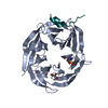
| ||||||||
|---|---|---|---|---|---|---|---|---|---|
| 1 |
| ||||||||
| Unit cell |
|
-Components
| #1: Protein | Mass: 39195.793 Da / Num. of mol.: 1 / Fragment: C-terminal WD40 Domain, residues 835-1186 Source method: isolated from a genetically manipulated source Source: (gene. exp.) Homo sapiens (human) / Gene: PALB2 / Plasmid: pTWO-B / Production host:   Spodoptera frugiperda (fall armyworm) / Strain (production host): SF9 / References: UniProt: Q86YC2 Spodoptera frugiperda (fall armyworm) / Strain (production host): SF9 / References: UniProt: Q86YC2 | ||
|---|---|---|---|
| #2: Protein/peptide | Mass: 2107.297 Da / Num. of mol.: 1 / Fragment: Interaction with PALB2, residues 21-39 / Source method: obtained synthetically Details: chemically synthesized; This sequence occurs naturally in humans References: UniProt: P51587 | ||
| #3: Chemical | Glycerol  Mass: 92.094 Da / Num. of mol.: 2 / Source method: obtained synthetically / Formula: C3H8O3 Mass: 92.094 Da / Num. of mol.: 2 / Source method: obtained synthetically / Formula: C3H8O3#4: Water | ChemComp-HOH / | Water Mass: 18.015 Da / Num. of mol.: 98 / Source method: isolated from a natural source / Formula: H2O Mass: 18.015 Da / Num. of mol.: 98 / Source method: isolated from a natural source / Formula: H2O |
-Experimental details
-Experiment
| Experiment | Method: X-RAY DIFFRACTION / Number of used crystals: 1 |
|---|
- Sample preparation
| Crystal | Density Matthews: 2.3 Å3/Da / Density % sol: 46.58 % |
|---|---|
| Crystal grow | Temperature: 293 K / Method: vapor diffusion, hanging drop / pH: 6.5 Details: 0.1M Na-Cacodylate pH 6.5, 0.2M magnesium acetate, 10% (w/v) PEG 8000, VAPOR DIFFUSION, HANGING DROP, temperature 293K |
-Data collection
| Diffraction | Mean temperature: 100 K |
|---|---|
| Diffraction source | Source: SYNCHROTRON / Site: ESRF  / Beamline: ID14-1 / Wavelength: 0.943 Å / Beamline: ID14-1 / Wavelength: 0.943 Å |
| Detector | Type: ADSC QUANTUM 210 / Detector: CCD / Date: Dec 8, 2007 / Details: Horizontally bended Ge(220) |
| Radiation | Monochromator: Diamond (111) / Protocol: SINGLE WAVELENGTH / Monochromatic (M) / Laue (L): M / Scattering type: x-ray |
| Radiation wavelength | Wavelength: 0.943 Å / Relative weight: 1 |
| Reflection | Resolution: 2.2→37.5 Å / Num. all: 19154 / Num. obs: 19026 / % possible obs: 99.9 % / Redundancy: 5.8 % / Biso Wilson estimate: 44.61 Å2 / Rmerge(I) obs: 0.07 / Rsym value: 0.07 / Net I/σ(I): 16.5 |
| Reflection shell | Resolution: 2.2→2.32 Å / Redundancy: 5.9 % / Rmerge(I) obs: 0.602 / Mean I/σ(I) obs: 2.9 / Num. unique all: 2800 / Rsym value: 0.602 / % possible all: 99 |
- Processing
| Software | Name: PHENIX / Version: (phenix.refine) / Classification: refinement | |||||||||||||||||||||||||||||||||||||||||||||||||||||||||||||||||||||||||||||||||||||||||||
|---|---|---|---|---|---|---|---|---|---|---|---|---|---|---|---|---|---|---|---|---|---|---|---|---|---|---|---|---|---|---|---|---|---|---|---|---|---|---|---|---|---|---|---|---|---|---|---|---|---|---|---|---|---|---|---|---|---|---|---|---|---|---|---|---|---|---|---|---|---|---|---|---|---|---|---|---|---|---|---|---|---|---|---|---|---|---|---|---|---|---|---|---|
| Refinement | Method to determine structure: MOLECULAR REPLACEMENT Starting model: PDB ENTRY 2w18 Resolution: 2.2→37.495 Å / Occupancy max: 1 / Occupancy min: 0.47 / FOM free R set: 0.802 / SU ML: 0.4 / Cross valid method: THROUGHOUT / σ(F): 0.91 / Phase error: 27.16 / Stereochemistry target values: ML
| |||||||||||||||||||||||||||||||||||||||||||||||||||||||||||||||||||||||||||||||||||||||||||
| Solvent computation | Shrinkage radii: 0.9 Å / VDW probe radii: 1.11 Å / Solvent model: FLAT BULK SOLVENT MODEL / Bsol: 60.519 Å2 / ksol: 0.37 e/Å3 | |||||||||||||||||||||||||||||||||||||||||||||||||||||||||||||||||||||||||||||||||||||||||||
| Displacement parameters | Biso max: 105.85 Å2 / Biso mean: 47.989 Å2 / Biso min: 23.46 Å2
| |||||||||||||||||||||||||||||||||||||||||||||||||||||||||||||||||||||||||||||||||||||||||||
| Refinement step | Cycle: LAST / Resolution: 2.2→37.495 Å
| |||||||||||||||||||||||||||||||||||||||||||||||||||||||||||||||||||||||||||||||||||||||||||
| Refine LS restraints |
| |||||||||||||||||||||||||||||||||||||||||||||||||||||||||||||||||||||||||||||||||||||||||||
| LS refinement shell | Refine-ID: X-RAY DIFFRACTION
|
