 Movie
Movie Controller

[English] 日本語
 Yorodumi
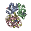
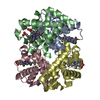
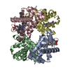
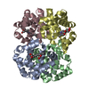
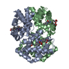
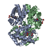
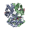
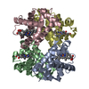
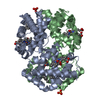
- PDB-3eu1: Crystal Structure determination of goat hemoglobin (Capra hircus)... -
+ Open data

- Basic information
| Entry | Database: PDB / ID: 3eu1 | ||||||
|---|---|---|---|---|---|---|---|
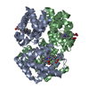
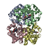
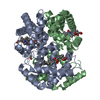
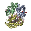
| Title | Crystal Structure determination of goat hemoglobin (Capra hircus) at 3 angstrom resolution | ||||||
 Components Components |
| ||||||
 Keywords Keywords |  Oxygen storage / Oxygen transport / low oxygen affinity / Capra hircus / hemoglobin / allosteric mechanism / 2 / 3-Diphospho glycerate Oxygen storage / Oxygen transport / low oxygen affinity / Capra hircus / hemoglobin / allosteric mechanism / 2 / 3-Diphospho glycerate | ||||||
| Function / homology |  Function and homology informationhaptoglobin binding / hemoglobin alpha binding / haptoglobin-hemoglobin complex / organic acid binding / hemoglobin complex / hydrogen peroxide catabolic process / oxygen carrier activity / peroxidase activity / oxygen binding / blood microparticle ...haptoglobin binding / hemoglobin alpha binding / haptoglobin-hemoglobin complex / organic acid binding / hemoglobin complex / hydrogen peroxide catabolic process / oxygen carrier activity / peroxidase activity / oxygen binding / blood microparticle / iron ion binding / heme binding / metal ion binding Function and homology informationhaptoglobin binding / hemoglobin alpha binding / haptoglobin-hemoglobin complex / organic acid binding / hemoglobin complex / hydrogen peroxide catabolic process / oxygen carrier activity / peroxidase activity / oxygen binding / blood microparticle ...haptoglobin binding / hemoglobin alpha binding / haptoglobin-hemoglobin complex / organic acid binding / hemoglobin complex / hydrogen peroxide catabolic process / oxygen carrier activity / peroxidase activity / oxygen binding / blood microparticle / iron ion binding / heme binding / metal ion bindingSimilarity search - Function | ||||||
| Biological species |  Capra hircus (goat) Capra hircus (goat) | ||||||
| Method | X-RAY DIFFRACTION / MOLECULAR REPLACEMENT / Resolution: 3 Å | ||||||
 Authors Authors | Sathya Moorthy, P. / Neelagandan, K. / Balasubramanian, M. / Ponnuswamy, M.N. | ||||||
 Citation Citation | Journal: Protein Pept.Lett. / Year: 2009 Title: Purification, crystallization and preliminary X-ray diffraction studies on goat (Capra hircus) hemoglobin - a low oxygen affinity species Authors: Sathya Moorthy, P. / Neelagandan, K. / Balasubramanian, M. / Ponnuswamy, M.N. | ||||||
| History |
|
- Structure visualization
| Structure viewer | Molecule: MolmilJmol/JSmol |
|---|
- Downloads & links
-Download
| PDBx/mmCIF format | 3eu1.cif.gz | 123 KB | Display | PDBx/mmCIF format |
|---|---|---|---|---|
| PDB format | pdb3eu1.ent.gz | 97.1 KB | Display | PDB format |
| PDBx/mmJSON format | 3eu1.json.gz | Tree view | PDBx/mmJSON format | |
| Others |  Other downloads Other downloads |
-Validation report
| Arichive directory | https://data.pdbj.org/pub/pdb/validation_reports/eu/3eu1ftp://data.pdbj.org/pub/pdb/validation_reports/eu/3eu1 | HTTPS FTP |
|---|
-Related structure data
| Related structure data |  3d1aS S: Starting model for refinement |
|---|---|
| Similar structure data |


-Links
 PDBj
PDBj


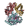
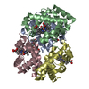
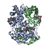
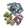
- Assembly
| Deposited unit | 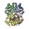
| ||||||||
|---|---|---|---|---|---|---|---|---|---|
| 1 |
| ||||||||
| Unit cell |
|
-Components
| #1: Protein | / Hemoglobin alpha-1/2 chain / Alpha-1/2-globin Mass: 15056.157 Da / Num. of mol.: 2 / Source method: isolated from a natural source / Source: (natural) Capra hircus (goat) / Cell: RED BLOOD CELLS / Tissue: blood / References: UniProt: P68238, UniProt: P0CH25*PLUS#2: Protein | / Hemoglobin beta-A chain / Beta-A-globin / Alanine beta-globinMass: 16046.405 Da / Num. of mol.: 2 / Source method: isolated from a natural source / Source: (natural) Capra hircus (goat) / Cell: RED BLOOD CELLS / Tissue: blood / References: UniProt: P02077#3: Chemical | ChemComp-HEM / Heme B  Mass: 616.487 Da / Num. of mol.: 4 / Source method: obtained synthetically / Formula: C34H32FeN4O4 Mass: 616.487 Da / Num. of mol.: 4 / Source method: obtained synthetically / Formula: C34H32FeN4O4 |
|---|
-Experimental details
-Experiment
| Experiment | Method: X-RAY DIFFRACTION / Number of used crystals: 1 |
|---|
- Sample preparation
| Crystal | Density Matthews: 2.27 Å3/Da / Density % sol: 45.92 % |
|---|---|
| Crystal grow | Temperature: 293 K / Method: vapor diffusion, hanging drop / pH: 7.5 Details: 2micro litres of Hb with 2micro litre of well solution containing 25mM of Phosphate buffer at pH 7.5 with 0.5M NaCl, VAPOR DIFFUSION, HANGING DROP, temperature 293K |
-Data collection
| Diffraction | Mean temperature: 293 K |
|---|---|
| Diffraction source | Source: ROTATING ANODE / Type: RIGAKU RUH3R / Wavelength: 1.5418 Å |
| Detector | Type: MAR scanner 345 mm plate / Detector: IMAGE PLATE / Date: Jul 16, 2008 |
| Radiation | Protocol: SINGLE WAVELENGTH / Monochromatic (M) / Laue (L): M / Scattering type: x-ray |
| Radiation wavelength | Wavelength: 1.5418 Å / Relative weight: 1 |
| Reflection | Resolution: 3→30 Å / Num. all: 10705 / Num. obs: 9607 / % possible obs: 89.7 % / Observed criterion σ(F): 3 / Observed criterion σ(I): 2 / Redundancy: 4.12 % / Rsym value: 0.1854 / Net I/σ(I): 2.9 |
| Reflection shell | Resolution: 3→3.11 Å / Redundancy: 3.55 % / Mean I/σ(I) obs: 0.7 / Rsym value: 0.5361 / % possible all: 93 |
- Processing
| Software |
| ||||||||||||||||||||||||||||||||||||||||||||||||||||||||||||||||||||||||||||||||||||||||||
|---|---|---|---|---|---|---|---|---|---|---|---|---|---|---|---|---|---|---|---|---|---|---|---|---|---|---|---|---|---|---|---|---|---|---|---|---|---|---|---|---|---|---|---|---|---|---|---|---|---|---|---|---|---|---|---|---|---|---|---|---|---|---|---|---|---|---|---|---|---|---|---|---|---|---|---|---|---|---|---|---|---|---|---|---|---|---|---|---|---|---|---|
| Refinement | Method to determine structure: MOLECULAR REPLACEMENT Starting model: PDB ENTRY 3d1a Resolution: 3→22.08 Å / Cor.coef. Fo:Fc: 0.907 / Cor.coef. Fo:Fc free: 0.835 / SU B: 22.731 / SU ML: 0.405 / Cross valid method: THROUGHOUT / ESU R Free: 0.617 / Stereochemistry target values: MAXIMUM LIKELIHOOD
| ||||||||||||||||||||||||||||||||||||||||||||||||||||||||||||||||||||||||||||||||||||||||||
| Solvent computation | Ion probe radii: 0.8 Å / Shrinkage radii: 0.8 Å / VDW probe radii: 1.4 Å / Solvent model: MASK | ||||||||||||||||||||||||||||||||||||||||||||||||||||||||||||||||||||||||||||||||||||||||||
| Displacement parameters | Biso max: 92.55 Å2 / Biso mean: 34.953 Å2 / Biso min: 3.44 Å2
| ||||||||||||||||||||||||||||||||||||||||||||||||||||||||||||||||||||||||||||||||||||||||||
| Refinement step | Cycle: LAST / Resolution: 3→22.08 Å
| ||||||||||||||||||||||||||||||||||||||||||||||||||||||||||||||||||||||||||||||||||||||||||
| Refine LS restraints |
| ||||||||||||||||||||||||||||||||||||||||||||||||||||||||||||||||||||||||||||||||||||||||||
| LS refinement shell | Resolution: 3.001→3.078 Å / Total num. of bins used: 20
|
