 Movie
Movie Controller


+ Open data

- Basic information
| Entry | Database: PDB / ID: 3eqp | ||||||
|---|---|---|---|---|---|---|---|
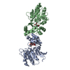
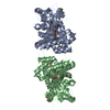
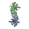
| Title | Crystal Structure of Ack1 with compound T95 | ||||||
 Components Components | Activated CDC42 kinase 1 | ||||||
 Keywords Keywords |  TRANSFERASE / ack1 / Alternative splicing / ATP-binding / Cell membrane / Kinase / Magnesium / Membrane / Metal-binding / Nucleotide-binding / Phosphoprotein / Polymorphism / SH3 domain / Tyrosine-protein kinase TRANSFERASE / ack1 / Alternative splicing / ATP-binding / Cell membrane / Kinase / Magnesium / Membrane / Metal-binding / Nucleotide-binding / Phosphoprotein / Polymorphism / SH3 domain / Tyrosine-protein kinase | ||||||
| Function / homology |  Function and homology informationregulation of clathrin-dependent endocytosis / cytoophidium / Grb2-EGFR complex / GTPase inhibitor activity / WW domain binding / clathrin-coated vesicle / epidermal growth factor receptor binding / small GTPase-mediated signal transduction / clathrin-coated pit / protein serine/threonine/tyrosine kinase activity ...regulation of clathrin-dependent endocytosis / cytoophidium / Grb2-EGFR complex / GTPase inhibitor activity / WW domain binding / clathrin-coated vesicle / epidermal growth factor receptor binding / small GTPase-mediated signal transduction / clathrin-coated pit / protein serine/threonine/tyrosine kinase activity / adherens junction / non-specific protein-tyrosine kinase / non-membrane spanning protein tyrosine kinase activity / cytoplasmic vesicle membrane / endocytosis / positive regulation of peptidyl-tyrosine phosphorylation / protein tyrosine kinase activity / cell surface receptor signaling pathway / non-specific serine/threonine protein kinase / endosome / phosphorylation / protein serine kinase activity / intracellular membrane-bounded organelle / protein serine/threonine kinase activity / ubiquitin protein ligase binding / perinuclear region of cytoplasm / ATP binding / membrane / identical protein binding / metal ion binding / nucleus / plasma membrane / cytosol / cytoplasm Function and homology informationregulation of clathrin-dependent endocytosis / cytoophidium / Grb2-EGFR complex / GTPase inhibitor activity / WW domain binding / clathrin-coated vesicle / epidermal growth factor receptor binding / small GTPase-mediated signal transduction / clathrin-coated pit / protein serine/threonine/tyrosine kinase activity ...regulation of clathrin-dependent endocytosis / cytoophidium / Grb2-EGFR complex / GTPase inhibitor activity / WW domain binding / clathrin-coated vesicle / epidermal growth factor receptor binding / small GTPase-mediated signal transduction / clathrin-coated pit / protein serine/threonine/tyrosine kinase activity / adherens junction / non-specific protein-tyrosine kinase / non-membrane spanning protein tyrosine kinase activity / cytoplasmic vesicle membrane / endocytosis / positive regulation of peptidyl-tyrosine phosphorylation / protein tyrosine kinase activity / cell surface receptor signaling pathway / non-specific serine/threonine protein kinase / endosome / phosphorylation / protein serine kinase activity / intracellular membrane-bounded organelle / protein serine/threonine kinase activity / ubiquitin protein ligase binding / perinuclear region of cytoplasm / ATP binding / membrane / identical protein binding / metal ion binding / nucleus / plasma membrane / cytosol / cytoplasmSimilarity search - Function | ||||||
| Biological species |  Homo sapiens (human) Homo sapiens (human) | ||||||
| Method | X-RAY DIFFRACTION / SYNCHROTRON / Resolution: 2.3 Å | ||||||
 Authors Authors | Liu, J. / Wang, Z. / Walker, N.P.C. | ||||||
 Citation Citation | Journal: Bioorg.Med.Chem.Lett. / Year: 2008 Title: Identification and optimization of N3,N6-diaryl-1H-pyrazolo[3,4-d]pyrimidine-3,6-diamines as a novel class of ACK1 inhibitors. Authors: Kopecky, D.J. / Hao, X. / Chen, Y. / Fu, J. / Jiao, X. / Jaen, J.C. / Cardozo, M.G. / Liu, J. / Wang, Z. / Walker, N.P. / Wesche, H. / Li, S. / Farrelly, E. / Xiao, S.H. / Kayser, F. | ||||||
| History |
|
- Structure visualization
| Structure viewer | Molecule: MolmilJmol/JSmol |
|---|
- Downloads & links
-Download
| PDBx/mmCIF format | 3eqp.cif.gz | 124 KB | Display | PDBx/mmCIF format |
|---|---|---|---|---|
| PDB format | pdb3eqp.ent.gz | 96.8 KB | Display | PDB format |
| PDBx/mmJSON format | 3eqp.json.gz | Tree view | PDBx/mmJSON format | |
| Others |  Other downloads Other downloads |
-Validation report
| Arichive directory | https://data.pdbj.org/pub/pdb/validation_reports/eq/3eqpftp://data.pdbj.org/pub/pdb/validation_reports/eq/3eqp | HTTPS FTP |
|---|
-Related structure data


-Links
 PDBj
PDBj
- Assembly
| Deposited unit | 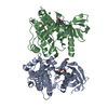
| ||||||||
|---|---|---|---|---|---|---|---|---|---|
| 1 | 
| ||||||||
| 2 | 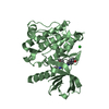
| ||||||||
| 3 | 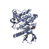
| ||||||||
| Unit cell |
|
-Components
| #1: Protein | Mass: 31538.395 Da / Num. of mol.: 2 / Fragment: UNP residues 117-392 Source method: isolated from a genetically manipulated source Source: (gene. exp.) Homo sapiens (human) / Gene: TNK2, ACK1 / Production host: Insect cellsReferences: UniProt: Q07912, non-specific protein-tyrosine kinase#2: Chemical |   Mass: 582.693 Da / Num. of mol.: 2 / Source method: obtained synthetically / Formula: C33H38N6O4 Mass: 582.693 Da / Num. of mol.: 2 / Source method: obtained synthetically / Formula: C33H38N6O4#3: Chemical | ChemComp-CL / | Chloride  Mass: 35.453 Da / Num. of mol.: 1 / Source method: obtained synthetically / Formula: Cl Mass: 35.453 Da / Num. of mol.: 1 / Source method: obtained synthetically / Formula: Cl#4: Water | ChemComp-HOH / | Water Mass: 18.015 Da / Num. of mol.: 172 / Source method: isolated from a natural source / Formula: H2O Mass: 18.015 Da / Num. of mol.: 172 / Source method: isolated from a natural source / Formula: H2O |
|---|
-Experimental details
-Experiment
| Experiment | Method: X-RAY DIFFRACTION / Number of used crystals: 1 |
|---|
- Sample preparation
| Crystal | Density Matthews: 2.22 Å3/Da / Density % sol: 44.58 % |
|---|---|
| Crystal grow | Temperature: 298 K / Method: vapor diffusion / Details: vapor diffusion, temperature 298K |
-Data collection
| Diffraction | Ambient temp details: 100 |
|---|---|
| Diffraction source | Source: SYNCHROTRON / Site: ALS  / Beamline: 5.0.2 / Wavelength: 1 Å / Beamline: 5.0.2 / Wavelength: 1 Å |
| Detector | Detector: CCD / Date: Sep 15, 2004 |
| Radiation | Protocol: SINGLE WAVELENGTH / Monochromatic (M) / Laue (L): M / Scattering type: x-ray |
| Radiation wavelength | Wavelength: 1 Å / Relative weight: 1 |
| Reflection | Resolution: 2.3→91.29 Å / Num. obs: 23616 / % possible obs: 94.9 % / Redundancy: 3.5 % / Rsym value: 0.11 / Net I/σ(I): 6.2 |
| Reflection shell | Resolution: 2.3→2.42 Å / % possible obs: 92 % / Redundancy: 3.5 % / Mean I/σ(I) obs: 1.5 / Rsym value: 0.485 |
- Processing
| Software |
| |||||||||||||||||||||||||||||||||||||||||||||||||||||||||||||||||
|---|---|---|---|---|---|---|---|---|---|---|---|---|---|---|---|---|---|---|---|---|---|---|---|---|---|---|---|---|---|---|---|---|---|---|---|---|---|---|---|---|---|---|---|---|---|---|---|---|---|---|---|---|---|---|---|---|---|---|---|---|---|---|---|---|---|---|
| Refinement | Resolution: 2.3→60.86 Å / Cor.coef. Fo:Fc: 0.915 / Cor.coef. Fo:Fc free: 0.854 / Occupancy max: 1 / Occupancy min: 1 / SU B: 8.686 / SU ML: 0.213 / Cross valid method: THROUGHOUT / σ(F): 0 / ESU R: 0.562 / ESU R Free: 0.306 / Stereochemistry target values: MAXIMUM LIKELIHOOD / Details: U VALUES: REFINED INDIVIDUALLY
| |||||||||||||||||||||||||||||||||||||||||||||||||||||||||||||||||
| Solvent computation | Ion probe radii: 0.8 Å / Shrinkage radii: 0.8 Å / VDW probe radii: 1.4 Å / Solvent model: BABINET MODEL WITH MASK | |||||||||||||||||||||||||||||||||||||||||||||||||||||||||||||||||
| Displacement parameters | Biso max: 61.6 Å2 / Biso mean: 27.266 Å2 / Biso min: 3.92 Å2
| |||||||||||||||||||||||||||||||||||||||||||||||||||||||||||||||||
| Refinement step | Cycle: LAST / Resolution: 2.3→60.86 Å
| |||||||||||||||||||||||||||||||||||||||||||||||||||||||||||||||||
| Refine LS restraints |
| |||||||||||||||||||||||||||||||||||||||||||||||||||||||||||||||||
| LS refinement shell | Resolution: 2.3→2.36 Å / Total num. of bins used: 20
|
