 Movie
Movie Controller


+ Open data

- Basic information
| Entry | Database: PDB / ID: 3efc | ||||||
|---|---|---|---|---|---|---|---|
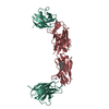
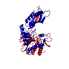
| Title | Crystal Structure of YaeT periplasmic domain | ||||||
 Components Components | Outer membrane protein assembly factor yaeT | ||||||
 Keywords Keywords |  MEMBRANE PROTEIN / POTRA fold / Cell membrane / Cell outer membrane / Membrane MEMBRANE PROTEIN / POTRA fold / Cell membrane / Cell outer membrane / Membrane | ||||||
| Function / homology |  Function and homology information Function and homology informationBam protein complex / Gram-negative-bacterium-type cell outer membrane assembly / protein insertion into membrane / cell outer membrane / cell adhesion / membraneSimilarity search - Function | ||||||
| Biological species |  Escherichia coli (E. coli) Escherichia coli (E. coli) | ||||||
| Method | X-RAY DIFFRACTION / SYNCHROTRON / MAD / Resolution: 3.3 Å | ||||||
 Authors Authors | Gatzeva-Topalova, P.Z. / Walton, T.A. / Sousa, M.C. | ||||||
 Citation Citation | Journal: Structure / Year: 2008 Title: Crystal structure of YaeT: conformational flexibility and substrate recognition. Authors: Gatzeva-Topalova, P.Z. / Walton, T.A. / Sousa, M.C. | ||||||
| History |
|
- Structure visualization
| Structure viewer | Molecule: MolmilJmol/JSmol |
|---|
- Downloads & links
-Download
| PDBx/mmCIF format | 3efc.cif.gz | 76.4 KB | Display | PDBx/mmCIF format |
|---|---|---|---|---|
| PDB format | pdb3efc.ent.gz | 56.5 KB | Display | PDB format |
| PDBx/mmJSON format | 3efc.json.gz | Tree view | PDBx/mmJSON format | |
| Others |  Other downloads Other downloads |
-Validation report
| Arichive directory | https://data.pdbj.org/pub/pdb/validation_reports/ef/3efcftp://data.pdbj.org/pub/pdb/validation_reports/ef/3efc | HTTPS FTP |
|---|
-Related structure data
| Similar structure data |
|---|


-Links
 PDBj
PDBj- Assembly
| Deposited unit | 
| ||||||||
|---|---|---|---|---|---|---|---|---|---|
| 1 |
| ||||||||
| Unit cell |
|
-Components
| #1: Protein | Mass: 44187.598 Da / Num. of mol.: 1 / Fragment: periplasmic domain (UNP residues 21-410) Source method: isolated from a genetically manipulated source Source: (gene. exp.) Escherichia coli (E. coli) / Gene: yaeT, yzzN, yzzY, b0177, JW0172 / Production host: Escherichia coli (E. coli) / Strain (production host): Rosetta (DE3) / References: UniProt: P0A940 |
|---|
-Experimental details
-Experiment
| Experiment | Method: X-RAY DIFFRACTION / Number of used crystals: 2 |
|---|
- Sample preparation
| Crystal | Density Matthews: 3.98 Å3/Da / Density % sol: 69.11 % |
|---|---|
| Crystal grow | Temperature: 289.15 K / Method: vapor diffusion, hanging drop / pH: 6.5 Details: 1.3-1.6M (NH4)2SO4, 3-6% polyethylene glycol 400, 10% Dioxane, 0.1M MES pH 6.5, VAPOR DIFFUSION, HANGING DROP, temperature 289.15K |
-Data collection
| Diffraction |
| |||||||||||||||
|---|---|---|---|---|---|---|---|---|---|---|---|---|---|---|---|---|
| Diffraction source |
| |||||||||||||||
| Detector |
| |||||||||||||||
| Radiation |
| |||||||||||||||
| Radiation wavelength |
| |||||||||||||||
| Reflection | Resolution: 3.3→50 Å / Num. obs: 11035 / % possible obs: 99.6 % / Redundancy: 3.5 % / Rsym value: 0.056 / Net I/σ(I): 20.6 | |||||||||||||||
| Reflection shell | Resolution: 3.3→3.42 Å / Redundancy: 3.6 % / Mean I/σ(I) obs: 2.3 / Num. unique all: 1069 / Rsym value: 0.424 / % possible all: 99.9 |

- Processing
| Software |
| |||||||||||||||||||||||||
|---|---|---|---|---|---|---|---|---|---|---|---|---|---|---|---|---|---|---|---|---|---|---|---|---|---|---|
| Refinement | Method to determine structure: MAD / Resolution: 3.3→47.5 Å / Cross valid method: THROUGHOUT / σ(F): 0
| |||||||||||||||||||||||||
| Displacement parameters | Biso mean: 95.5 Å2 | |||||||||||||||||||||||||
| Refinement step | Cycle: LAST / Resolution: 3.3→47.5 Å
| |||||||||||||||||||||||||
| LS refinement shell | Resolution: 3.3→3.45 Å
|
