 Movie
Movie Controller

[English] 日本語
 Yorodumi
- PDB-3eci: Microtubule-associated protein 1 light chain 3 alpha isoform A (M... -
+ Open data

- Basic information
| Entry | Database: PDB / ID: 3eci | ||||||
|---|---|---|---|---|---|---|---|
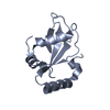
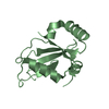
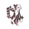
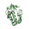
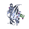
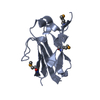
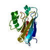
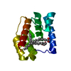
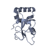
| Title | Microtubule-associated protein 1 light chain 3 alpha isoform A (MAP1ALC3) | ||||||
 Components Components | Microtubule-associated protein 1 light chain 3 alpha | ||||||
 Keywords Keywords |  APOPTOSIS / UBIQUITIN-LIKE (UB ROLL) / AUTOPHAGY / CYTOPLASM / CYTOPLASMIC VESICLE / LIPOPROTEIN / MEMBRANE / MICROTUBULE / UBL CONJUGATION PATHWAY / STRUCTURAL GENOMICS CONSORTIUM / Alternative splicing APOPTOSIS / UBIQUITIN-LIKE (UB ROLL) / AUTOPHAGY / CYTOPLASM / CYTOPLASMIC VESICLE / LIPOPROTEIN / MEMBRANE / MICROTUBULE / UBL CONJUGATION PATHWAY / STRUCTURAL GENOMICS CONSORTIUM / Alternative splicing | ||||||
| Function / homology |  Function and homology information Function and homology informationcellular response to oxygen-glucose deprivation / autophagy of mitochondrion / cellular response to nitrogen starvation / SMAD protein signal transduction / response to iron(II) ion / autolysosome / Macroautophagy / Receptor Mediated Mitophagy / p38MAPK cascade / autophagosome maturation ...cellular response to oxygen-glucose deprivation / autophagy of mitochondrion / cellular response to nitrogen starvation / SMAD protein signal transduction / response to iron(II) ion / autolysosome / Macroautophagy / Receptor Mediated Mitophagy / p38MAPK cascade / autophagosome maturation / autophagosome membrane / organelle membrane / autophagosome assembly / autophagosome / JNK cascade / cellular response to copper ion / cellular response to amino acid starvation / PINK1-PRKN Mediated Mitophagy / cellular response to starvation / macroautophagy / response to lead ion / phospholipid binding / cellular response to hydrogen peroxide / late endosome / microtubule binding / microtubule / intracellular membrane-bounded organelle / glutamatergic synapse / ubiquitin protein ligase binding / cytosolSimilarity search - Function | ||||||
| Biological species |  Homo sapiens (human) Homo sapiens (human) | ||||||
| Method | X-RAY DIFFRACTION / SYNCHROTRON / MOLECULAR REPLACEMENT / Resolution: 2.65 Å | ||||||
 Authors Authors | Walker, J.R. / Davis, T.L. / Mujib, S. / Butler-Cole, C. / Tempel, W. / Weigelt, J. / Bountra, C. / Arrowsmith, C.H. / Edwards, A.M. / Botchkarev, A. / Dhe-Paganon, S. | ||||||
 Citation Citation | Journal: To be Published Title: Human Autophagy-Related Protein LC3 A Authors: Walker, J.R. / Davis, T.L. / Mujib, S. / Butler-Cole, C. / Tempel, W. / Bountra, C. / Weigelt, J. / Arrowsmith, C.H. / Edwards, A.M. / Bochkarev, A. / Dhe-Paganon, S. | ||||||
| History |
|
- Structure visualization
| Structure viewer | Molecule: MolmilJmol/JSmol |
|---|
- Downloads & links
-Download
| PDBx/mmCIF format | 3eci.cif.gz | 97.3 KB | Display | PDBx/mmCIF format |
|---|---|---|---|---|
| PDB format | pdb3eci.ent.gz | 75.2 KB | Display | PDB format |
| PDBx/mmJSON format | 3eci.json.gz | Tree view | PDBx/mmJSON format | |
| Others |  Other downloads Other downloads |
-Validation report
| Arichive directory | https://data.pdbj.org/pub/pdb/validation_reports/ec/3eciftp://data.pdbj.org/pub/pdb/validation_reports/ec/3eci | HTTPS FTP |
|---|
-Related structure data
| Related structure data | 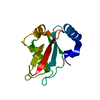 1ugmS S: Starting model for refinement |
|---|---|
| Similar structure data |

-Links
 PDBj
PDBj


- Assembly
| Deposited unit | 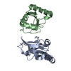
| ||||||||
|---|---|---|---|---|---|---|---|---|---|
| 1 | 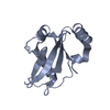
| ||||||||
| 2 | 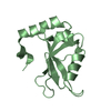
| ||||||||
| Unit cell |
|
-Components
| #1: Protein | Mass: 14350.503 Da / Num. of mol.: 2 Source method: isolated from a genetically manipulated source Details: Isoform A (MAPALC3) / Source: (gene. exp.) Homo sapiens (human) / Gene: MAP1LC3A / Plasmid: PET28-MHL / Production host:  Escherichia coli (E. coli) / Strain (production host): BL21 (DE3) Gold / References: UniProt: Q9H492 Escherichia coli (E. coli) / Strain (production host): BL21 (DE3) Gold / References: UniProt: Q9H492 |
|---|
-Experimental details
-Experiment
| Experiment | Method: X-RAY DIFFRACTION / Number of used crystals: 1 |
|---|
- Sample preparation
| Crystal | Density Matthews: 2.07 Å3/Da / Density % sol: 40.7 % |
|---|---|
| Crystal grow | Temperature: 298 K / Method: vapor diffusion, hanging drop / pH: 4.5 Details: 32 % PEG 4000, 0.1 M NA ACETATE, PH 4.50, 0.2 M AMMONIUM ACETATE, VAPOR DIFFUSION, HANGING DROP, CRYOPROTECTION 20% GLYCEROL, TEMPERATURE 298K |
-Data collection
| Diffraction | Mean temperature: 100 K |
|---|---|
| Diffraction source | Source: SYNCHROTRON / Site: APS  / Beamline: 23-ID-D / Wavelength: 0.97926 / Wavelength: 0.97926 Å / Beamline: 23-ID-D / Wavelength: 0.97926 / Wavelength: 0.97926 Å |
| Detector | Type: MARMOSAIC 300 mm CCD / Detector: CCD / Date: Apr 15, 2007 / Details: MIRRORS |
| Radiation | Monochromator: DOUBLE CRYSTAL / Protocol: SINGLE WAVELENGTH / Monochromatic (M) / Laue (L): M / Scattering type: x-ray |
| Radiation wavelength | Wavelength: 0.97926 Å / Relative weight: 1 |
| Reflection | Resolution: 2.65→40 Å / Num. all: 7058 / Num. obs: 7058 / % possible obs: 99.4 % / Observed criterion σ(F): 0 / Observed criterion σ(I): -3 / Redundancy: 4.1 % / Rsym value: 0.059 |
| Reflection shell | Resolution: 2.65→2.74 Å / Redundancy: 4.1 % / Mean I/σ(I) obs: 2.19 / Num. unique all: 682 / Rsym value: 0.713 / % possible all: 100 |
- Processing
| Software |
| |||||||||||||||||||||||||||||||||||||||||||||||||||||||||||||||||||||||||||||||||||||
|---|---|---|---|---|---|---|---|---|---|---|---|---|---|---|---|---|---|---|---|---|---|---|---|---|---|---|---|---|---|---|---|---|---|---|---|---|---|---|---|---|---|---|---|---|---|---|---|---|---|---|---|---|---|---|---|---|---|---|---|---|---|---|---|---|---|---|---|---|---|---|---|---|---|---|---|---|---|---|---|---|---|---|---|---|---|---|
| Refinement | Method to determine structure: MOLECULAR REPLACEMENT Starting model: PDB entry 1UGM Resolution: 2.65→28.31 Å / Cor.coef. Fo:Fc: 0.943 / Cor.coef. Fo:Fc free: 0.909 / SU B: 41.44 / SU ML: 0.37 / Cross valid method: THROUGHOUT / ESU R: 10.469 / ESU R Free: 0.411 / Stereochemistry target values: MAXIMUM LIKELIHOOD Details: HYDROGENS HAVE BEEN ADDED IN THE RIDING POSITIONS. ATOM RECORD CONTAINS SUM OF TLS AND RESIDUAL B FACTORS. ANISOU RECORD CONTAINS SUM OF TLS AND RESIDUAL U FACTORS.
| |||||||||||||||||||||||||||||||||||||||||||||||||||||||||||||||||||||||||||||||||||||
| Solvent computation | Ion probe radii: 0.8 Å / Shrinkage radii: 0.8 Å / VDW probe radii: 1.2 Å / Solvent model: MASK | |||||||||||||||||||||||||||||||||||||||||||||||||||||||||||||||||||||||||||||||||||||
| Displacement parameters | Biso mean: 37.478 Å2
| |||||||||||||||||||||||||||||||||||||||||||||||||||||||||||||||||||||||||||||||||||||
| Refinement step | Cycle: LAST / Resolution: 2.65→28.31 Å
| |||||||||||||||||||||||||||||||||||||||||||||||||||||||||||||||||||||||||||||||||||||
| Refine LS restraints |
| |||||||||||||||||||||||||||||||||||||||||||||||||||||||||||||||||||||||||||||||||||||
| LS refinement shell | Resolution: 2.65→2.718 Å / Total num. of bins used: 20
| |||||||||||||||||||||||||||||||||||||||||||||||||||||||||||||||||||||||||||||||||||||
| Refinement TLS params. | Method: refined / Refine-ID: X-RAY DIFFRACTION
| |||||||||||||||||||||||||||||||||||||||||||||||||||||||||||||||||||||||||||||||||||||
| Refinement TLS group |
|
