 Movie
Movie Controller


+ Open data

- Basic information
| Entry | Database: PDB / ID: 3e8g | ||||||
|---|---|---|---|---|---|---|---|
| Title | Crystal Structure of the the open NaK channel-Na+/Ca2+ complex | ||||||
 Components Components | Potassium channel protein | ||||||
 Keywords Keywords | MEMBRANE PROTEIN / non-selective cation channel / tetrameric cation channel family / 2-transmembrane helix channels / Ionic channel | ||||||
| Function / homology |  Function and homology information Function and homology informationstabilization of membrane potential / potassium ion leak channel activity / outward rectifier potassium channel activity / membrane / identical protein binding / metal ion bindingSimilarity search - Function | ||||||
| Biological species |  Bacillus cereus (bacteria) Bacillus cereus (bacteria) | ||||||
| Method | X-RAY DIFFRACTION / SYNCHROTRON / MOLECULAR REPLACEMENT / molecular replacement / Resolution: 2 Å | ||||||
 Authors Authors | Jiang, Y. / Alam, A. | ||||||
 Citation Citation | Journal: Nat.Struct.Mol.Biol. / Year: 2009 Title: Structural analysis of ion selectivity in the NaK channel Authors: Alam, A. / Jiang, Y. | ||||||
| History |
|
- Structure visualization
| Structure viewer | Molecule: MolmilJmol/JSmol |
|---|
- Downloads & links
-Download
| PDBx/mmCIF format | 3e8g.cif.gz | 50.5 KB | Display | PDBx/mmCIF format |
|---|---|---|---|---|
| PDB format | pdb3e8g.ent.gz | 36.8 KB | Display | PDB format |
| PDBx/mmJSON format | 3e8g.json.gz | Tree view | PDBx/mmJSON format | |
| Others |  Other downloads Other downloads |
-Validation report
| Arichive directory | https://data.pdbj.org/pub/pdb/validation_reports/e8/3e8gftp://data.pdbj.org/pub/pdb/validation_reports/e8/3e8g | HTTPS FTP |
|---|
-Related structure data


-Links
 PDBj
PDBj

- Assembly
| Deposited unit | 
| |||||||||||||||||||||||||||||||||
|---|---|---|---|---|---|---|---|---|---|---|---|---|---|---|---|---|---|---|---|---|---|---|---|---|---|---|---|---|---|---|---|---|---|---|
| 1 | 
| |||||||||||||||||||||||||||||||||
| 2 | 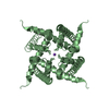
| |||||||||||||||||||||||||||||||||
| Unit cell |
| |||||||||||||||||||||||||||||||||
| Components on special symmetry positions |
|
-Components
-Protein , 1 types, 2 molecules AB
| #1: Protein | Mass: 10706.538 Da / Num. of mol.: 2 / Fragment: transmembrane domain, residues 19-110 Source method: isolated from a genetically manipulated source Source: (gene. exp.) Bacillus cereus (bacteria) / Plasmid: pQE60 / Production host: Escherichia coli (E. coli) / Strain (production host): SG13009 / References: UniProt: Q81HW2 |
|---|
-Non-polymers , 5 types, 63 molecules 


| #2: Chemical | 2-Methyl-2,4-pentanediol Mass: 118.174 Da / Num. of mol.: 3 / Source method: obtained synthetically / Formula: C6H14O2 / Comment: precipitant*YM Mass: 118.174 Da / Num. of mol.: 3 / Source method: obtained synthetically / Formula: C6H14O2 / Comment: precipitant*YM#3: Chemical |  Mass: 132.905 Da / Num. of mol.: 2 / Source method: obtained synthetically / Formula: Cs Mass: 132.905 Da / Num. of mol.: 2 / Source method: obtained synthetically / Formula: Cs#4: Chemical |  Mass: 40.078 Da / Num. of mol.: 2 / Source method: obtained synthetically / Formula: Ca Mass: 40.078 Da / Num. of mol.: 2 / Source method: obtained synthetically / Formula: Ca#5: Chemical | ChemComp-NA /  Mass: 22.990 Da / Num. of mol.: 4 / Source method: obtained synthetically / Formula: Na Mass: 22.990 Da / Num. of mol.: 4 / Source method: obtained synthetically / Formula: Na#6: Water | ChemComp-HOH / | WaterMass: 18.015 Da / Num. of mol.: 52 / Source method: isolated from a natural source / Formula: H2O |
|---|
-Experimental details
-Experiment
| Experiment | Method: X-RAY DIFFRACTION / Number of used crystals: 1 |
|---|
- Sample preparation
| Crystal | Density Matthews: 2.41 Å3/Da / Density % sol: 48.88 % |
|---|---|
| Crystal grow | Temperature: 293 K / Method: vapor diffusion, sitting drop / pH: 7.5 Details: 100mM Hepes, 55-70% (4S)-2-Methyl-2,4-pentanediol (MPD), 10mM CaCl2, pH 7.5, VAPOR DIFFUSION, SITTING DROP, temperature 293K |
-Data collection
| Diffraction | Mean temperature: 100 K |
|---|---|
| Diffraction source | Source: SYNCHROTRON / Site: APS  / Beamline: 19-ID / Beamline: 19-ID |
| Detector | Type: ADSC QUANTUM 315 / Detector: CCD |
| Radiation | Protocol: SINGLE WAVELENGTH / Monochromatic (M) / Laue (L): M / Scattering type: x-ray |
| Radiation wavelength | Relative weight: 1 |
| Reflection | Resolution: 2→50 Å / Num. obs: 13398 |
-Phasing
| Phasing | Method: molecular replacement |
|---|
- Processing
| Software |
| ||||||||||||||||||||||||
|---|---|---|---|---|---|---|---|---|---|---|---|---|---|---|---|---|---|---|---|---|---|---|---|---|---|
| Refinement | Method to determine structure: MOLECULAR REPLACEMENT / Resolution: 2→33.99 Å / Occupancy max: 1 / Occupancy min: 0.5 / σ(F): 0
| ||||||||||||||||||||||||
| Solvent computation | Bsol: 113.782 Å2 | ||||||||||||||||||||||||
| Displacement parameters | Biso max: 101.3 Å2 / Biso mean: 50.48 Å2 / Biso min: 23.96 Å2
| ||||||||||||||||||||||||
| Refinement step | Cycle: LAST / Resolution: 2→33.99 Å
| ||||||||||||||||||||||||
| Refine LS restraints |
| ||||||||||||||||||||||||
| Xplor file |
|
