 Movie
Movie Controller

[English] 日本語
 Yorodumi
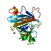
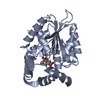
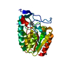
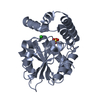
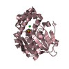
- PDB-3e0x: The crystal structure of a Lipase-esterase related protein from C... -
+ Open data

- Basic information
| Entry | Database: PDB / ID: 3e0x | ||||||
|---|---|---|---|---|---|---|---|
| Title | The crystal structure of a Lipase-esterase related protein from Clostridium acetobutylicum ATCC 824 | ||||||
 Components Components | Lipase-esterase related protein | ||||||
 Keywords Keywords |  structural genomics / unknown function / APC60309 / Lipase-esterase related protein / Clostridium acetobutylicum ATCC 824 / PSI-2 / midwest center for structural genomics / MCSG / Protein Structure Initiative structural genomics / unknown function / APC60309 / Lipase-esterase related protein / Clostridium acetobutylicum ATCC 824 / PSI-2 / midwest center for structural genomics / MCSG / Protein Structure Initiative | ||||||
| Function / homology | Serine aminopeptidase, S33 / Serine aminopeptidase, S33 / Alpha/Beta hydrolase fold, catalytic domain / Alpha/Beta hydrolase fold / Rossmann fold / 3-Layer(aba) Sandwich / Alpha Beta / ORTHO-XYLENE / Lipase-esterase related protein Function and homology information Function and homology information | ||||||
| Biological species |  Clostridium acetobutylicum (bacteria) Clostridium acetobutylicum (bacteria) | ||||||
| Method | X-RAY DIFFRACTION / SYNCHROTRON / SAD / Resolution: 1.45 Å | ||||||
 Authors Authors | Tan, K. / Sather, A. / Cobb, G. / Joachimiak, A. / Midwest Center for Structural Genomics (MCSG) | ||||||
 Citation Citation | Journal: To be Published Title: The crystal structure of a Lipase-esterase related protein from Clostridium acetobutylicum ATCC 824 Authors: Tan, K. / Sather, A. / Cobb, G. / Joachimiak, A. | ||||||
| History |
|
- Structure visualization
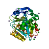
| Structure viewer | Molecule: MolmilJmol/JSmol |
|---|
- Downloads & links
-Download
| PDBx/mmCIF format | 3e0x.cif.gz | 229.2 KB | Display | PDBx/mmCIF format |
|---|---|---|---|---|
| PDB format | pdb3e0x.ent.gz | 193.3 KB | Display | PDB format |
| PDBx/mmJSON format | 3e0x.json.gz | Tree view | PDBx/mmJSON format | |
| Others |  Other downloads Other downloads |
-Validation report
| Arichive directory | https://data.pdbj.org/pub/pdb/validation_reports/e0/3e0xftp://data.pdbj.org/pub/pdb/validation_reports/e0/3e0x | HTTPS FTP |
|---|
-Related structure data
| Similar structure data | |
|---|---|
| Other databases |


-Links
 PDBj
PDBj- Assembly
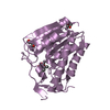
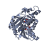
| Deposited unit | 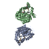
| ||||||||
|---|---|---|---|---|---|---|---|---|---|
| 1 | 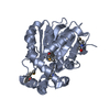
| ||||||||
| 2 | 
| ||||||||
| Unit cell |
| ||||||||
| Details | Authors state that the biological assembly is experimentally unknown. From molecular packing, the molecule is expected to be a monomer. |
-Components
| #1: Protein | Mass: 27630.303 Da / Num. of mol.: 2 Source method: isolated from a genetically manipulated source Source: (gene. exp.) Clostridium acetobutylicum (bacteria) / Strain: ATCC 824 / Gene: CA_C0816 / Plasmid: pMCSG7 / Production host: Escherichia coli (E. coli) / Strain (production host): BL21 / References: UniProt: Q97KV0#2: Chemical | O-Xylene  Mass: 106.165 Da / Num. of mol.: 2 / Source method: obtained synthetically / Formula: C8H10 Mass: 106.165 Da / Num. of mol.: 2 / Source method: obtained synthetically / Formula: C8H10#3: Water | ChemComp-HOH / | Water Mass: 18.015 Da / Num. of mol.: 754 / Source method: isolated from a natural source / Formula: H2O Mass: 18.015 Da / Num. of mol.: 754 / Source method: isolated from a natural source / Formula: H2O |
|---|
-Experimental details
-Experiment
| Experiment | Method: X-RAY DIFFRACTION / Number of used crystals: 1 |
|---|
- Sample preparation
| Crystal | Density Matthews: 2.05 Å3/Da / Density % sol: 39.98 % |
|---|---|
| Crystal grow | Temperature: 289 K / Method: vapor diffusion, sitting drop Details: 0.2M LiCl 20% PEG3350, VAPOR DIFFUSION, SITTING DROP, temperature 289K |
-Data collection
| Diffraction | Mean temperature: 100 K |
|---|---|
| Diffraction source | Source: SYNCHROTRON / Site: APS  / Beamline: 19-ID / Wavelength: 0.97929 Å / Beamline: 19-ID / Wavelength: 0.97929 Å |
| Detector | Type: ADSC QUANTUM 315 / Detector: CCD / Date: Apr 1, 2008 / Details: Mirror |
| Radiation | Monochromator: Si crystal 111 / Protocol: SINGLE WAVELENGTH / Monochromatic (M) / Laue (L): M / Scattering type: x-ray |
| Radiation wavelength | Wavelength: 0.97929 Å / Relative weight: 1 |
| Reflection | Resolution: 1.45→38.1 Å / Num. all: 75965 / Num. obs: 75965 / % possible obs: 96.3 % / Observed criterion σ(F): 0 / Observed criterion σ(I): 0 / Redundancy: 4.2 % / Rmerge(I) obs: 0.092 / Net I/σ(I): 28.9 |
| Reflection shell | Resolution: 1.45→1.48 Å / Redundancy: 2.8 % / Rmerge(I) obs: 0.49 / Mean I/σ(I) obs: 2.28 / Num. unique all: 2968 / % possible all: 75.8 |
- Processing
| Software |
| ||||||||||||||||||||||||||||||||||||||||||||||||||||||||||||||||||||||||||||||||||||||||||||||||||||||||||||||||||||||||||||||||||||||||||||||||||||||||||||||||||||||||||
|---|---|---|---|---|---|---|---|---|---|---|---|---|---|---|---|---|---|---|---|---|---|---|---|---|---|---|---|---|---|---|---|---|---|---|---|---|---|---|---|---|---|---|---|---|---|---|---|---|---|---|---|---|---|---|---|---|---|---|---|---|---|---|---|---|---|---|---|---|---|---|---|---|---|---|---|---|---|---|---|---|---|---|---|---|---|---|---|---|---|---|---|---|---|---|---|---|---|---|---|---|---|---|---|---|---|---|---|---|---|---|---|---|---|---|---|---|---|---|---|---|---|---|---|---|---|---|---|---|---|---|---|---|---|---|---|---|---|---|---|---|---|---|---|---|---|---|---|---|---|---|---|---|---|---|---|---|---|---|---|---|---|---|---|---|---|---|---|---|---|---|---|
| Refinement | Method to determine structure: SAD / Resolution: 1.45→38.1 Å / Cor.coef. Fo:Fc: 0.973 / Cor.coef. Fo:Fc free: 0.958 / SU B: 2.292 / SU ML: 0.041 / Cross valid method: THROUGHOUT / σ(F): 0 / σ(I): 0 / ESU R: 0.087 / ESU R Free: 0.071 / Stereochemistry target values: MAXIMUM LIKELIHOOD / Details: HYDROGENS HAVE BEEN ADDED IN THE RIDING POSITIONS
| ||||||||||||||||||||||||||||||||||||||||||||||||||||||||||||||||||||||||||||||||||||||||||||||||||||||||||||||||||||||||||||||||||||||||||||||||||||||||||||||||||||||||||
| Solvent computation | Ion probe radii: 0.8 Å / Shrinkage radii: 0.8 Å / VDW probe radii: 1.2 Å / Solvent model: MASK | ||||||||||||||||||||||||||||||||||||||||||||||||||||||||||||||||||||||||||||||||||||||||||||||||||||||||||||||||||||||||||||||||||||||||||||||||||||||||||||||||||||||||||
| Displacement parameters | Biso mean: 15.368 Å2
| ||||||||||||||||||||||||||||||||||||||||||||||||||||||||||||||||||||||||||||||||||||||||||||||||||||||||||||||||||||||||||||||||||||||||||||||||||||||||||||||||||||||||||
| Refinement step | Cycle: LAST / Resolution: 1.45→38.1 Å
| ||||||||||||||||||||||||||||||||||||||||||||||||||||||||||||||||||||||||||||||||||||||||||||||||||||||||||||||||||||||||||||||||||||||||||||||||||||||||||||||||||||||||||
| Refine LS restraints |
| ||||||||||||||||||||||||||||||||||||||||||||||||||||||||||||||||||||||||||||||||||||||||||||||||||||||||||||||||||||||||||||||||||||||||||||||||||||||||||||||||||||||||||
| LS refinement shell | Resolution: 1.45→1.488 Å / Total num. of bins used: 20
|
