 Movie
Movie Controller


+ Open data

- Basic information
| Entry | Database: PDB / ID: 300000000 | ||||||
|---|---|---|---|---|---|---|---|
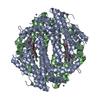
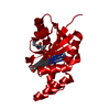
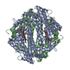
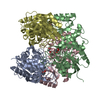
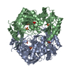
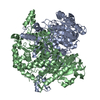
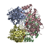
| Title | H55S mutant Xanthomonas campestris tryptophan 2,3-dioxygenase | ||||||
 Components Components | Tryptophan 2,3-dioxygenase | ||||||
 Keywords Keywords | OXIDOREDUCTASE / TDO / heme / tryptophan 2 / 3-dioxygenase / H55S / Dioxygenase | ||||||
| Function / homology |  Function and homology information Function and homology informationtryptophan catabolic process to acetyl-CoA / tryptophan 2,3-dioxygenase / tryptophan 2,3-dioxygenase activity / tryptophan catabolic process to kynurenine / heme binding / metal ion bindingSimilarity search - Function | ||||||
| Biological species |  Xanthomonas campestris pv. campestris (bacteria) Xanthomonas campestris pv. campestris (bacteria) | ||||||
| Method | X-RAY DIFFRACTION / SYNCHROTRON / MOLECULAR REPLACEMENT / Resolution: 1.9 Å | ||||||
 Authors Authors | Mowat, C.G. / Campbell, L.P. | ||||||
 Citation Citation | Journal: Biochemistry / Year: 2008 Title: Histidine 55 of tryptophan 2,3-dioxygenase is not an active site base but regulates catalysis by controlling substrate binding Authors: Thackray, S.J. / Bruckmann, C. / Anderson, J.L. / Campbell, L.P. / Xiao, R. / Zhao, L. / Mowat, C.G. / Forouhar, F. / Tong, L. / Chapman, S.K. | ||||||
| History |
|
- Structure visualization
| Structure viewer | Molecule: MolmilJmol/JSmol |
|---|
- Downloads & links
-Download
| PDBx/mmCIF format | 3e08.cif.gz | 508.5 KB | Display | PDBx/mmCIF format |
|---|---|---|---|---|
| PDB format | pdb3e08.ent.gz | 416.6 KB | Display | PDB format |
| PDBx/mmJSON format | 3e08.json.gz | Tree view | PDBx/mmJSON format | |
| Others |  Other downloads Other downloads |
-Validation report
| Arichive directory | https://data.pdbj.org/pub/pdb/validation_reports/e0/3e08ftp://data.pdbj.org/pub/pdb/validation_reports/e0/3e08 | HTTPS FTP |
|---|
-Related structure data
| Related structure data | 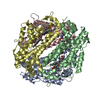 3bk9C  2nw7S C: citing same article ( S: Starting model for refinement |
|---|---|
| Similar structure data |


-Links
 PDBj
PDBj
- Assembly
| Deposited unit | 
| ||||||||
|---|---|---|---|---|---|---|---|---|---|
| 1 | 
| ||||||||
| 2 | 
| ||||||||
| Unit cell |
|
-Components
| #1: Protein | Mass: 34609.215 Da / Num. of mol.: 8 / Mutation: H55S Source method: isolated from a genetically manipulated source Source: (gene. exp.) Xanthomonas campestris pv. campestris (bacteria)Gene: xccb100_0466 / Plasmid: pMGK / Production host: Escherichia coli (E. coli) / Strain (production host): BL21 (DE3) / References: UniProt: Q8PDA8, tryptophan 2,3-dioxygenase#2: Chemical | ChemComp-TRP / Tryptophan  Type: L-peptide linking / Mass: 204.225 Da / Num. of mol.: 16 / Source method: obtained synthetically / Formula: C11H12N2O2 Type: L-peptide linking / Mass: 204.225 Da / Num. of mol.: 16 / Source method: obtained synthetically / Formula: C11H12N2O2#3: Chemical | ChemComp-HEM / Heme B  Mass: 616.487 Da / Num. of mol.: 8 / Source method: obtained synthetically / Formula: C34H32FeN4O4 Mass: 616.487 Da / Num. of mol.: 8 / Source method: obtained synthetically / Formula: C34H32FeN4O4#4: Water | ChemComp-HOH / | Water Mass: 18.015 Da / Num. of mol.: 1955 / Source method: isolated from a natural source / Formula: H2O Mass: 18.015 Da / Num. of mol.: 1955 / Source method: isolated from a natural source / Formula: H2O |
|---|
-Experimental details
-Experiment
| Experiment | Method: X-RAY DIFFRACTION / Number of used crystals: 1 |
|---|
- Sample preparation
| Crystal | Density Matthews: 2.29 Å3/Da / Density % sol: 46.3 % |
|---|---|
| Crystal grow | Temperature: 291 K / Method: vapor diffusion, hanging drop / pH: 6.3 Details: 9-10% PEG1000, 80mM MES (pH6.3), 20mM bicine (pH9.0), 40mM MnCl2, 400mM MgCl2, 8-15mM NaCN, 20mM L-Trp, VAPOR DIFFUSION, HANGING DROP, temperature 291K |
-Data collection
| Diffraction | Mean temperature: 100 K |
|---|---|
| Diffraction source | Source: SYNCHROTRON / Site: SRS  / Beamline: PX10.1 / Wavelength: 1.045 Å / Beamline: PX10.1 / Wavelength: 1.045 Å |
| Detector | Type: MAR CCD 165 mm / Detector: CCD / Date: Oct 10, 2007 |
| Radiation | Protocol: SINGLE WAVELENGTH / Monochromatic (M) / Laue (L): M / Scattering type: x-ray |
| Radiation wavelength | Wavelength: 1.045 Å / Relative weight: 1 |
| Reflection | Resolution: 1.9→17.56 Å / Num. obs: 189203 / % possible obs: 93.6 % / Observed criterion σ(F): 0 / Observed criterion σ(I): 0 / Redundancy: 4.1 % / Biso Wilson estimate: 23.24 Å2 / Rmerge(I) obs: 0.078 / Net I/σ(I): 7.3 |
| Reflection shell | Resolution: 1.9→2 Å / Redundancy: 3.8 % / Rmerge(I) obs: 0.48 / Mean I/σ(I) obs: 2.6 / Num. unique all: 24898 / % possible all: 87.1 |
- Processing
| Software |
| |||||||||||||||||||||||||
|---|---|---|---|---|---|---|---|---|---|---|---|---|---|---|---|---|---|---|---|---|---|---|---|---|---|---|
| Refinement | Method to determine structure: MOLECULAR REPLACEMENT Starting model: PDB ENTRY 2NW7 Resolution: 1.9→17.56 Å / Cross valid method: THROUGHOUT / σ(F): 0 / σ(I): 0 / Stereochemistry target values: Engh & Huber
| |||||||||||||||||||||||||
| Displacement parameters | Biso mean: 23.2 Å2 | |||||||||||||||||||||||||
| Refinement step | Cycle: LAST / Resolution: 1.9→17.56 Å
| |||||||||||||||||||||||||
| Refine LS restraints |
| |||||||||||||||||||||||||
| LS refinement shell | Resolution: 1.9→1.9216 Å
|
