 Movie
Movie Controller

[English] 日本語
 Yorodumi
- PDB-3dwg: Crystal structure of a sulfur carrier protein complex found in th... -
+ Open data

- Basic information
| Entry | Database: PDB / ID: 3dwg | ||||||
|---|---|---|---|---|---|---|---|
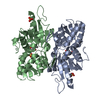
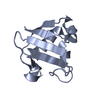
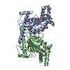
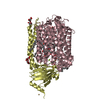
| Title | Crystal structure of a sulfur carrier protein complex found in the cysteine biosynthetic pathway of Mycobacterium tuberculosis | ||||||
 Components Components |
| ||||||
 Keywords Keywords |  TRANSFERASE / Sulfur carrier protein complex / beta-grasp fold / Amino-acid biosynthesis / Cysteine biosynthesis / Pyridoxal phosphate TRANSFERASE / Sulfur carrier protein complex / beta-grasp fold / Amino-acid biosynthesis / Cysteine biosynthesis / Pyridoxal phosphate | ||||||
| Function / homology |  Function and homology information Function and homology information[CysO sulfur-carrier protein]-thiocarboxylate-dependent cysteine synthase / Cysteine synthesis from O-phosphoserine / O-phosphoserine sulfhydrylase activity / cysteine biosynthetic process / cysteine synthase activity / L-cysteine desulfhydrase activity / cysteine biosynthetic process from serine / pyridoxal phosphate binding / protein-containing complex / cytosol / cytoplasmSimilarity search - Function | ||||||
| Biological species |   Mycobacterium tuberculosis (bacteria) Mycobacterium tuberculosis (bacteria) | ||||||
| Method | X-RAY DIFFRACTION / SYNCHROTRON / SAD / Resolution: 1.53 Å | ||||||
 Authors Authors | Jurgenson, C.T. / Burns, K.E. / Begley, T.P. / Ealick, S.E. | ||||||
 Citation Citation | Journal: Biochemistry / Year: 2008 Title: Crystal structure of a sulfur carrier protein complex found in the cysteine biosynthetic pathway of Mycobacterium tuberculosis. Authors: Jurgenson, C.T. / Burns, K.E. / Begley, T.P. / Ealick, S.E. | ||||||
| History |
|
- Structure visualization
| Structure viewer | Molecule: MolmilJmol/JSmol |
|---|
- Downloads & links
-Download
| PDBx/mmCIF format | 3dwg.cif.gz | 167.6 KB | Display | PDBx/mmCIF format |
|---|---|---|---|---|
| PDB format | pdb3dwg.ent.gz | 136.5 KB | Display | PDB format |
| PDBx/mmJSON format | 3dwg.json.gz | Tree view | PDBx/mmJSON format | |
| Others |  Other downloads Other downloads |
-Validation report
| Arichive directory | https://data.pdbj.org/pub/pdb/validation_reports/dw/3dwgftp://data.pdbj.org/pub/pdb/validation_reports/dw/3dwg | HTTPS FTP |
|---|
-Related structure data


-Links
 PDBj
PDBj


- Assembly
| Deposited unit | 
| ||||||||
|---|---|---|---|---|---|---|---|---|---|
| 1 |
| ||||||||
| Unit cell |
| ||||||||
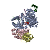
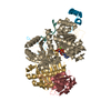
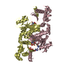
| Details | The asymmetric unit contains the biological unit consisting of two CysM protomers and one CysO protomer. |
-Components
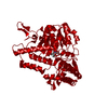
| #1: Protein | Mass: 34767.207 Da / Num. of mol.: 2 Source method: isolated from a genetically manipulated source Source: (gene. exp.) Mycobacterium tuberculosis (bacteria) / Strain: H37Rv / Gene: cysM, Rv1336, MT1377, MTCY130.21 / Plasmid: pET16b / Production host: Escherichia coli (E. coli) / Strain (production host): BL21(DE3)B834References: UniProt: P63873, UniProt: P9WP53*PLUS, cysteine synthase#2: Protein | | Mass: 9564.656 Da / Num. of mol.: 1 Source method: isolated from a genetically manipulated source Source: (gene. exp.) Mycobacterium tuberculosis (bacteria) / Strain: H37Rv / Gene: cfp10A, Rv1335, MT1376.1, MTCY130.20 / Plasmid: pET16b / Production host: Escherichia coli (E. coli) / Strain (production host): BL21(DE3) / References: UniProt: P0A646, UniProt: P9WP33*PLUS#3: Chemical | Pyridoxal phosphate  Mass: 247.142 Da / Num. of mol.: 2 / Source method: obtained synthetically / Formula: C8H10NO6P Mass: 247.142 Da / Num. of mol.: 2 / Source method: obtained synthetically / Formula: C8H10NO6P#4: Water | ChemComp-HOH / | Water Mass: 18.015 Da / Num. of mol.: 985 / Source method: isolated from a natural source / Formula: H2O Mass: 18.015 Da / Num. of mol.: 985 / Source method: isolated from a natural source / Formula: H2O |
|---|
-Experimental details
-Experiment
| Experiment | Method: X-RAY DIFFRACTION / Number of used crystals: 2 |
|---|
- Sample preparation
| Crystal | Density Matthews: 2.45 Å3/Da / Density % sol: 49.74 % |
|---|---|
| Crystal grow | Temperature: 295 K / pH: 5.8 Details: 7-10% w/v PEG 4000, 0.1 M Sodium citrate pH 5.8, 0.2 M Ammonium acetate, VAPOR DIFFUSION, HANGING DROP, temperature 295K, pH 5.80 |
-Data collection
| Diffraction |
| ||||||||||||||||||
|---|---|---|---|---|---|---|---|---|---|---|---|---|---|---|---|---|---|---|---|
| Diffraction source |
| ||||||||||||||||||
| Detector |
| ||||||||||||||||||
| Radiation |
| ||||||||||||||||||
| Radiation wavelength | Wavelength: 0.9795 Å / Relative weight: 1 | ||||||||||||||||||
| Reflection | Resolution: 1.53→50 Å / Num. obs: 105098 / % possible obs: 96.3 % / Redundancy: 3.18 % / Rsym value: 0.046 / Net I/σ(I): 26.2 | ||||||||||||||||||
| Reflection shell | Resolution: 1.53→1.57 Å / Redundancy: 2.5 % / Mean I/σ(I) obs: 4 / Rsym value: 0.219 / % possible all: 81.6 |

- Processing
| Software |
| ||||||||||||||||||||||||||||||||||||||||||||||||||||||||||||||||||||||||||||||||||||||||||||||||||||||||||||||||||||||||||||||||||||||||||||||||||||||||||||||||||||||||||
|---|---|---|---|---|---|---|---|---|---|---|---|---|---|---|---|---|---|---|---|---|---|---|---|---|---|---|---|---|---|---|---|---|---|---|---|---|---|---|---|---|---|---|---|---|---|---|---|---|---|---|---|---|---|---|---|---|---|---|---|---|---|---|---|---|---|---|---|---|---|---|---|---|---|---|---|---|---|---|---|---|---|---|---|---|---|---|---|---|---|---|---|---|---|---|---|---|---|---|---|---|---|---|---|---|---|---|---|---|---|---|---|---|---|---|---|---|---|---|---|---|---|---|---|---|---|---|---|---|---|---|---|---|---|---|---|---|---|---|---|---|---|---|---|---|---|---|---|---|---|---|---|---|---|---|---|---|---|---|---|---|---|---|---|---|---|---|---|---|---|---|---|
| Refinement | Method to determine structure: SAD / Resolution: 1.53→27.13 Å / Cor.coef. Fo:Fc: 0.966 / Cor.coef. Fo:Fc free: 0.946 / SU B: 1.481 / SU ML: 0.055 / Cross valid method: THROUGHOUT / ESU R: 0.08 / ESU R Free: 0.085 / Stereochemistry target values: MAXIMUM LIKELIHOOD / Details: HYDROGENS HAVE BEEN ADDED IN THE RIDING POSITIONS
| ||||||||||||||||||||||||||||||||||||||||||||||||||||||||||||||||||||||||||||||||||||||||||||||||||||||||||||||||||||||||||||||||||||||||||||||||||||||||||||||||||||||||||
| Solvent computation | Ion probe radii: 0.8 Å / Shrinkage radii: 0.8 Å / VDW probe radii: 1.2 Å / Solvent model: MASK | ||||||||||||||||||||||||||||||||||||||||||||||||||||||||||||||||||||||||||||||||||||||||||||||||||||||||||||||||||||||||||||||||||||||||||||||||||||||||||||||||||||||||||
| Displacement parameters | Biso mean: 26.69 Å2
| ||||||||||||||||||||||||||||||||||||||||||||||||||||||||||||||||||||||||||||||||||||||||||||||||||||||||||||||||||||||||||||||||||||||||||||||||||||||||||||||||||||||||||
| Refinement step | Cycle: LAST / Resolution: 1.53→27.13 Å
| ||||||||||||||||||||||||||||||||||||||||||||||||||||||||||||||||||||||||||||||||||||||||||||||||||||||||||||||||||||||||||||||||||||||||||||||||||||||||||||||||||||||||||
| Refine LS restraints |
| ||||||||||||||||||||||||||||||||||||||||||||||||||||||||||||||||||||||||||||||||||||||||||||||||||||||||||||||||||||||||||||||||||||||||||||||||||||||||||||||||||||||||||
| LS refinement shell | Resolution: 1.53→1.57 Å / Total num. of bins used: 20
|
