 Movie
Movie Controller


+ Open data

- Basic information
| Entry | Database: PDB / ID: 3dwd | ||||||
|---|---|---|---|---|---|---|---|
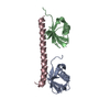
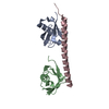
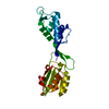
| Title | Crystal structure of the ArfGAP domain of human ARFGAP1 | ||||||
 Components Components | ADP-ribosylation factor GTPase-activating protein 1 | ||||||
 Keywords Keywords |  TRANSPORT PROTEIN / GAP / GTPASE ACTIVATING PROTEIN / STRUCTURAL GENOMICS CONSORTIUM (SGC) / ER-Golgi transport / Golgi apparatus / GTPase activation / Metal-binding / Phosphoprotein / Protein transport / Transport / Zinc-finger TRANSPORT PROTEIN / GAP / GTPASE ACTIVATING PROTEIN / STRUCTURAL GENOMICS CONSORTIUM (SGC) / ER-Golgi transport / Golgi apparatus / GTPase activation / Metal-binding / Phosphoprotein / Protein transport / Transport / Zinc-finger | ||||||
| Function / homology |  Function and homology information Function and homology informationXBP1(S) activates chaperone genes / regulation of ARF protein signal transduction / COPI-dependent Golgi-to-ER retrograde traffic / regulation of endocytosis / COPI-mediated anterograde transport / vesicle-mediated transport / GTPase activator activity / protein transport / Clathrin-mediated endocytosis / Golgi membrane ...XBP1(S) activates chaperone genes / regulation of ARF protein signal transduction / COPI-dependent Golgi-to-ER retrograde traffic / regulation of endocytosis / COPI-mediated anterograde transport / vesicle-mediated transport / GTPase activator activity / protein transport / Clathrin-mediated endocytosis / Golgi membrane / synapse / metal ion binding / cytosolSimilarity search - Function | ||||||
| Biological species |  Homo sapiens (human) Homo sapiens (human) | ||||||
| Method | X-RAY DIFFRACTION / SYNCHROTRON / MOLECULAR REPLACEMENT / molecular replacement / Resolution: 2.4 Å | ||||||
 Authors Authors | Nedyalkova, L. / Tong, Y. / Tempel, W. / Landry, R. / Arrowsmith, C.H. / Edwards, A.M. / Bountra, C. / Wilkstrom, M. / Bochkarev, A. / Park, H. / Structural Genomics Consortium (SGC) | ||||||
 Citation Citation | Journal: To be Published Title: Crystal structure of the ArfGAP domain of human ARFGAP1 Authors: Nedyalkova, L. / Tong, Y. / Tempel, W. / Landry, R. / Arrowsmith, C.H. / Edwards, A.M. / Bountra, C. / Wilkstrom, M. / Bochkarev, A. / Park, H. | ||||||
| History |
|
- Structure visualization
| Structure viewer | Molecule: MolmilJmol/JSmol |
|---|
- Downloads & links
-Download
| PDBx/mmCIF format | 3dwd.cif.gz | 60.8 KB | Display | PDBx/mmCIF format |
|---|---|---|---|---|
| PDB format | pdb3dwd.ent.gz | 41.7 KB | Display | PDB format |
| PDBx/mmJSON format | 3dwd.json.gz | Tree view | PDBx/mmJSON format | |
| Others |  Other downloads Other downloads |
-Validation report
| Arichive directory | https://data.pdbj.org/pub/pdb/validation_reports/dw/3dwdftp://data.pdbj.org/pub/pdb/validation_reports/dw/3dwd | HTTPS FTP |
|---|
-Related structure data
| Related structure data |  2gf9S S: Starting model for refinement |
|---|---|
| Similar structure data |


-Links
 PDBj
PDBj


- Assembly
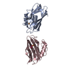
| Deposited unit | 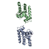
| ||||||||
|---|---|---|---|---|---|---|---|---|---|
| 1 |
| ||||||||
| 2 |
| ||||||||
| Unit cell |
| ||||||||
| Details | THE AUTHORS STATE THAT THE BIOLOGICAL UNIT OF THIS MACROMOLECULE IS UNKNOWN. |
-Components
| #1: Protein | Mass: 16850.082 Da / Num. of mol.: 2 / Fragment: Arf-GAP domain, UNP residues 1-128 Source method: isolated from a genetically manipulated source Source: (gene. exp.) Homo sapiens (human) / Gene: ARFGAP1, ARF1GAP / Plasmid: pET28a-LIC (GI:145307000) / Production host:  Escherichia coli (E. coli) / Strain (production host): BL21-CodonPlus(DE3)-V2R / References: UniProt: Q8N6T3 Escherichia coli (E. coli) / Strain (production host): BL21-CodonPlus(DE3)-V2R / References: UniProt: Q8N6T3#2: Chemical |   Mass: 65.409 Da / Num. of mol.: 2 / Source method: obtained synthetically / Formula: Zn Mass: 65.409 Da / Num. of mol.: 2 / Source method: obtained synthetically / Formula: Zn#3: Chemical |   Num. of mol.: 2 / Source method: obtained synthetically Num. of mol.: 2 / Source method: obtained synthetically#4: Water | ChemComp-HOH / | Water Mass: 18.015 Da / Num. of mol.: 11 / Source method: isolated from a natural source / Formula: H2O Mass: 18.015 Da / Num. of mol.: 11 / Source method: isolated from a natural source / Formula: H2O |
|---|
-Experimental details
-Experiment
| Experiment | Method: X-RAY DIFFRACTION / Number of used crystals: 1 |
|---|
- Sample preparation
| Crystal | Density Matthews: 2.36 Å3/Da / Density % sol: 47.81 % |
|---|---|
| Crystal grow | Temperature: 291 K / Method: vapor diffusion, sitting drop / pH: 8.5 Details: 3.5M Sodium Formate, 0.1M Tris, 1:100 chymotrypsin, pH 8.5, vapor diffusion, sitting drop, temperature 291K |
-Data collection
| Diffraction | Mean temperature: 100 K | |||||||||||||||||||||||||||||||||||||||||||||||||||||||||||||||||||||||||||||||||||||||||||||||||||||||||||||||||||||||||||||||||||||||||||||||||||
|---|---|---|---|---|---|---|---|---|---|---|---|---|---|---|---|---|---|---|---|---|---|---|---|---|---|---|---|---|---|---|---|---|---|---|---|---|---|---|---|---|---|---|---|---|---|---|---|---|---|---|---|---|---|---|---|---|---|---|---|---|---|---|---|---|---|---|---|---|---|---|---|---|---|---|---|---|---|---|---|---|---|---|---|---|---|---|---|---|---|---|---|---|---|---|---|---|---|---|---|---|---|---|---|---|---|---|---|---|---|---|---|---|---|---|---|---|---|---|---|---|---|---|---|---|---|---|---|---|---|---|---|---|---|---|---|---|---|---|---|---|---|---|---|---|---|---|---|---|
| Diffraction source | Source: SYNCHROTRON / Site: APS  / Beamline: 19-ID / Wavelength: 0.97935 Å / Beamline: 19-ID / Wavelength: 0.97935 Å | |||||||||||||||||||||||||||||||||||||||||||||||||||||||||||||||||||||||||||||||||||||||||||||||||||||||||||||||||||||||||||||||||||||||||||||||||||
| Detector | Type: ADSC QUANTUM 315 / Detector: CCD / Date: Jul 9, 2008 | |||||||||||||||||||||||||||||||||||||||||||||||||||||||||||||||||||||||||||||||||||||||||||||||||||||||||||||||||||||||||||||||||||||||||||||||||||
| Radiation | Protocol: SINGLE WAVELENGTH / Monochromatic (M) / Laue (L): M / Scattering type: x-ray | |||||||||||||||||||||||||||||||||||||||||||||||||||||||||||||||||||||||||||||||||||||||||||||||||||||||||||||||||||||||||||||||||||||||||||||||||||
| Radiation wavelength | Wavelength: 0.97935 Å / Relative weight: 1 | |||||||||||||||||||||||||||||||||||||||||||||||||||||||||||||||||||||||||||||||||||||||||||||||||||||||||||||||||||||||||||||||||||||||||||||||||||
| Reflection | Resolution: 2.4→50 Å / Num. obs: 12298 / % possible obs: 99.6 % / Redundancy: 7.4 % / Rmerge(I) obs: 0.092 / Χ2: 1.3 / Net I/σ(I): 8.1 | |||||||||||||||||||||||||||||||||||||||||||||||||||||||||||||||||||||||||||||||||||||||||||||||||||||||||||||||||||||||||||||||||||||||||||||||||||
| Reflection shell |
|
-Phasing
| Phasing | Method: molecular replacement | |||||||||
|---|---|---|---|---|---|---|---|---|---|---|
| Phasing MR | Model details: Phaser MODE: MR_AUTO
|
- Processing
| Software |
| |||||||||||||||||||||||||||||||||||||||||||||||||||||||||||||||||||||||||||||||||||||||||||||||||||||||||||||||||||||||||||||||||||||||||||||||||||
|---|---|---|---|---|---|---|---|---|---|---|---|---|---|---|---|---|---|---|---|---|---|---|---|---|---|---|---|---|---|---|---|---|---|---|---|---|---|---|---|---|---|---|---|---|---|---|---|---|---|---|---|---|---|---|---|---|---|---|---|---|---|---|---|---|---|---|---|---|---|---|---|---|---|---|---|---|---|---|---|---|---|---|---|---|---|---|---|---|---|---|---|---|---|---|---|---|---|---|---|---|---|---|---|---|---|---|---|---|---|---|---|---|---|---|---|---|---|---|---|---|---|---|---|---|---|---|---|---|---|---|---|---|---|---|---|---|---|---|---|---|---|---|---|---|---|---|---|---|
| Refinement | Method to determine structure: MOLECULAR REPLACEMENT Starting model: pdb entry 2gf9 Resolution: 2.4→30 Å / Cor.coef. Fo:Fc: 0.929 / Cor.coef. Fo:Fc free: 0.899 / WRfactor Rfree: 0.261 / WRfactor Rwork: 0.222 / SU B: 23.85 / SU ML: 0.244 / TLS residual ADP flag: LIKELY RESIDUAL / Cross valid method: THROUGHOUT / σ(F): 0 / ESU R: 0.354 / ESU R Free: 0.259 / Stereochemistry target values: MAXIMUM LIKELIHOOD Details: ATOM RECORD CONTAINS RESIDUAL B FACTORS ONLY. HYDROGENS HAVE BEEN ADDED IN THE RIDING POSITIONS U VALUES : RESIDUAL ONLY
| |||||||||||||||||||||||||||||||||||||||||||||||||||||||||||||||||||||||||||||||||||||||||||||||||||||||||||||||||||||||||||||||||||||||||||||||||||
| Solvent computation | Ion probe radii: 0.8 Å / Shrinkage radii: 0.8 Å / VDW probe radii: 1.2 Å / Solvent model: MASK BULK SOLVENT | |||||||||||||||||||||||||||||||||||||||||||||||||||||||||||||||||||||||||||||||||||||||||||||||||||||||||||||||||||||||||||||||||||||||||||||||||||
| Displacement parameters | Biso mean: 21.648 Å2
| |||||||||||||||||||||||||||||||||||||||||||||||||||||||||||||||||||||||||||||||||||||||||||||||||||||||||||||||||||||||||||||||||||||||||||||||||||
| Refinement step | Cycle: LAST / Resolution: 2.4→30 Å
| |||||||||||||||||||||||||||||||||||||||||||||||||||||||||||||||||||||||||||||||||||||||||||||||||||||||||||||||||||||||||||||||||||||||||||||||||||
| Refine LS restraints |
| |||||||||||||||||||||||||||||||||||||||||||||||||||||||||||||||||||||||||||||||||||||||||||||||||||||||||||||||||||||||||||||||||||||||||||||||||||
| LS refinement shell | Refine-ID: X-RAY DIFFRACTION / Total num. of bins used: 20
| |||||||||||||||||||||||||||||||||||||||||||||||||||||||||||||||||||||||||||||||||||||||||||||||||||||||||||||||||||||||||||||||||||||||||||||||||||
| Refinement TLS params. | Method: refined / Refine-ID: X-RAY DIFFRACTION
| |||||||||||||||||||||||||||||||||||||||||||||||||||||||||||||||||||||||||||||||||||||||||||||||||||||||||||||||||||||||||||||||||||||||||||||||||||
| Refinement TLS group |
|
