 Movie
Movie Controller

[English] 日本語
 Yorodumi
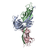
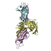
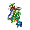
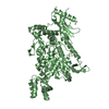
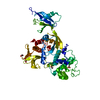
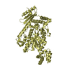
- PDB-3dpb: Crystal structure of the complex of the Caf1M chaperone with the ... -
+ Open data

- Basic information
| Entry | Database: PDB / ID: 3dpb | ||||||
|---|---|---|---|---|---|---|---|
| Title | Crystal structure of the complex of the Caf1M chaperone with the mini-fiber of two Caf1 subunits (Caf1:Caf1), carrying the Ala9Val, Ala11Val, and Leu13Val mutations in the Gd donor strand | ||||||
 Components Components |
| ||||||
 Keywords Keywords | CHAPERONE/STRUCTURAL PROTEIN / DONOR STRAND COMPLEMENTATION / FIMBRIAE /  CHAPERONE / PROTEIN-PROTEIN COMPLEX / BETA BARREL / Immunoglobulin domain / Periplasm / Plasmid / Capsule / Secreted / CHAPERONE-STRUCTURAL PROTEIN COMPLEX CHAPERONE / PROTEIN-PROTEIN COMPLEX / BETA BARREL / Immunoglobulin domain / Periplasm / Plasmid / Capsule / Secreted / CHAPERONE-STRUCTURAL PROTEIN COMPLEX | ||||||
| Function / homology |  Function and homology information Function and homology informationcapsule / pilus / chaperone-mediated protein folding / cell wall organization / outer membrane-bounded periplasmic space / cell adhesion / extracellular regionSimilarity search - Function | ||||||
| Biological species |   Yersinia pestis (bacteria) Yersinia pestis (bacteria) | ||||||
| Method | X-RAY DIFFRACTION / SYNCHROTRON / MOLECULAR REPLACEMENT / Resolution: 2.2 Å | ||||||
 Authors Authors | Fooks, L.J. / Yu, X. / Moslehi-Mohebi, E. / Tischenko, V. / Knight, S.D. / MacIntyre, S. / Zavialov, A.V. | ||||||
 Citation Citation | Journal: To be published Title: Hydrophobicity and rigidity of binding segments enable CAF1M chaperone to act as assembly catalyst Authors: Fooks, L.J. / Yu, X. / Moslehi-Mohebi, E. / Tischenko, V. / Knight, S.D. / MacIntyre, S. / Zavialov, A.V. | ||||||
| History |
|
- Structure visualization
| Structure viewer | Molecule: MolmilJmol/JSmol |
|---|
- Downloads & links
-Download
| PDBx/mmCIF format | 3dpb.cif.gz | 104.4 KB | Display | PDBx/mmCIF format |
|---|---|---|---|---|
| PDB format | pdb3dpb.ent.gz | 79.7 KB | Display | PDB format |
| PDBx/mmJSON format | 3dpb.json.gz | Tree view | PDBx/mmJSON format | |
| Others |  Other downloads Other downloads |
-Validation report
| Arichive directory | https://data.pdbj.org/pub/pdb/validation_reports/dp/3dpbftp://data.pdbj.org/pub/pdb/validation_reports/dp/3dpb | HTTPS FTP |
|---|
-Related structure data
| Related structure data | 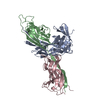 3dosC  3dsnC  1z9sS S: Starting model for refinement C: citing same article ( |
|---|---|
| Similar structure data |

-Links
 PDBj
PDBj
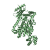
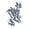
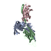
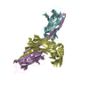
- Assembly
| Deposited unit | 
| ||||||||
|---|---|---|---|---|---|---|---|---|---|
| 1 |
| ||||||||
| Unit cell |
|
-Components
| #1: Protein | / CAPSULE PROTEIN FRACTION 1 MACHINERY Mass: 26329.012 Da / Num. of mol.: 1 / Fragment: UNP residues 24 to 258 Source method: isolated from a genetically manipulated source Source: (gene. exp.) Yersinia pestis (bacteria) / Gene: caf1M, YPMT1.82, y5194, y1098, YP_pMT084 / Production host: Escherichia coli (E. coli) / References: UniProt: P26926 | ||
|---|---|---|---|
| #2: Protein | Mass: 15617.234 Da / Num. of mol.: 2 / Fragment: UNP residues22 to 170 / Mutation: Ala9Val, Ala11Val, Leu13Val Source method: isolated from a genetically manipulated source Source: (gene. exp.) Yersinia pestis (bacteria) / Gene: caf1, YPMT1.84, y5196, y1100, YP_pMT082 / Production host: Escherichia coli (E. coli) / References: UniProt: P26948#3: Water | ChemComp-HOH / | Water Mass: 18.015 Da / Num. of mol.: 130 / Source method: isolated from a natural source / Formula: H2O Mass: 18.015 Da / Num. of mol.: 130 / Source method: isolated from a natural source / Formula: H2O |
-Experimental details
-Experiment
| Experiment | Method: X-RAY DIFFRACTION / Number of used crystals: 1 |
|---|
- Sample preparation
| Crystal | Density Matthews: 2.49 Å3/Da / Density % sol: 50.65 % |
|---|---|
| Crystal grow | Temperature: 295 K / Method: vapor diffusion, hanging drop / pH: 5.9 Details: 16-17% PEG 8000 in 0.1 M Na cacodylate and 0.2 M Ca acetate, pH 5.9, VAPOR DIFFUSION, HANGING DROP, temperature 295K |
-Data collection
| Diffraction | Mean temperature: 100 K |
|---|---|
| Diffraction source | Source: SYNCHROTRON / Site: ESRF  / Beamline: ID14-2 / Wavelength: 0.933 Å / Beamline: ID14-2 / Wavelength: 0.933 Å |
| Detector | Type: ADSC QUANTUM 4 / Detector: CCD / Date: Feb 27, 2006 |
| Radiation | Monochromator: Diamond (111), Ge(220) / Protocol: SINGLE WAVELENGTH / Monochromatic (M) / Laue (L): M / Scattering type: x-ray |
| Radiation wavelength | Wavelength: 0.933 Å / Relative weight: 1 |
| Reflection | Resolution: 2.2→40 Å / Num. obs: 24702 / % possible obs: 82.1 % / Observed criterion σ(F): 2 / Observed criterion σ(I): 2 / Redundancy: 4.6 % / Biso Wilson estimate: 20.85 Å2 / Rmerge(I) obs: 0.065 / Net I/σ(I): 20.8 |
| Reflection shell | Resolution: 2.2→2.32 Å / % possible all: 72.4 |
- Processing
| Software |
| ||||||||||||||||||||||||||||||||||||||||||||||||||||||||||||||||||||||||||||||||||||||||||
|---|---|---|---|---|---|---|---|---|---|---|---|---|---|---|---|---|---|---|---|---|---|---|---|---|---|---|---|---|---|---|---|---|---|---|---|---|---|---|---|---|---|---|---|---|---|---|---|---|---|---|---|---|---|---|---|---|---|---|---|---|---|---|---|---|---|---|---|---|---|---|---|---|---|---|---|---|---|---|---|---|---|---|---|---|---|---|---|---|---|---|---|
| Refinement | Method to determine structure: MOLECULAR REPLACEMENT Starting model: PDB entry 1Z9S Resolution: 2.2→40 Å / Cor.coef. Fo:Fc: 0.93 / Cor.coef. Fo:Fc free: 0.904 / SU B: 5.207 / SU ML: 0.136 / Cross valid method: THROUGHOUT / σ(F): 2 / ESU R: 0.312 / ESU R Free: 0.232 / Stereochemistry target values: MAXIMUM LIKELIHOOD / Details: HYDROGENS HAVE BEEN ADDED IN THE RIDING POSITIONS
| ||||||||||||||||||||||||||||||||||||||||||||||||||||||||||||||||||||||||||||||||||||||||||
| Solvent computation | Ion probe radii: 0.8 Å / Shrinkage radii: 0.8 Å / VDW probe radii: 1.2 Å / Solvent model: MASK | ||||||||||||||||||||||||||||||||||||||||||||||||||||||||||||||||||||||||||||||||||||||||||
| Displacement parameters | Biso mean: 20.85 Å2
| ||||||||||||||||||||||||||||||||||||||||||||||||||||||||||||||||||||||||||||||||||||||||||
| Refinement step | Cycle: LAST / Resolution: 2.2→40 Å
| ||||||||||||||||||||||||||||||||||||||||||||||||||||||||||||||||||||||||||||||||||||||||||
| Refine LS restraints |
| ||||||||||||||||||||||||||||||||||||||||||||||||||||||||||||||||||||||||||||||||||||||||||
| LS refinement shell | Resolution: 2.2→2.257 Å / Total num. of bins used: 20
|
