 Movie
Movie Controller

[English] 日本語
 Yorodumi
- PDB-3dmw: Crystal structure of human type III collagen G982-G1023 containin... -
+ Open data

- Basic information
| Entry | Database: PDB / ID: 3dmw | ||||||
|---|---|---|---|---|---|---|---|
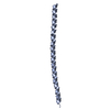
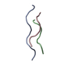
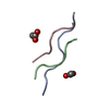
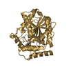
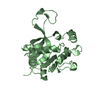
| Title | Crystal structure of human type III collagen G982-G1023 containing C-terminal cystine knot | ||||||
 Components Components | Collagen alpha-1(III) chain | ||||||
 Keywords Keywords |  STRUCTURAL PROTEIN / collagen III / cystine knot / triple helix / glycine / MAD phasing / Alternative splicing / Disease mutation / Ehlers-Danlos syndrome / Extracellular matrix / Glycoprotein / Hydroxylation / Phosphoprotein / Polymorphism / Secreted STRUCTURAL PROTEIN / collagen III / cystine knot / triple helix / glycine / MAD phasing / Alternative splicing / Disease mutation / Ehlers-Danlos syndrome / Extracellular matrix / Glycoprotein / Hydroxylation / Phosphoprotein / Polymorphism / Secreted | ||||||
| Function / homology |  Function and homology information Function and homology informationcollagen type III trimer / aorta smooth muscle tissue morphogenesis / limb joint morphogenesis / transforming growth factor beta1 production / elastic fiber assembly / negative regulation of neuron migration / Collagen chain trimerization / platelet-derived growth factor binding / endochondral bone morphogenesis / extracellular matrix structural constituent conferring tensile strength ...collagen type III trimer / aorta smooth muscle tissue morphogenesis / limb joint morphogenesis / transforming growth factor beta1 production / elastic fiber assembly / negative regulation of neuron migration / Collagen chain trimerization / platelet-derived growth factor binding / endochondral bone morphogenesis / extracellular matrix structural constituent conferring tensile strength / basement membrane organization / Extracellular matrix organization / layer formation in cerebral cortex / Collagen biosynthesis and modifying enzymes / peptide cross-linking / tissue homeostasis / Signaling by PDGF / negative regulation of immune response / NCAM1 interactions / digestive tract development / response to angiotensin / collagen fibril organization / Scavenging by Class A Receptors / extracellular matrix structural constituent / skin development / MET activates PTK2 signaling / Assembly of collagen fibrils and other multimeric structures / Syndecan interactions / positive regulation of Rho protein signal transduction / SMAD binding / Collagen degradation / Non-integrin membrane-ECM interactions / ECM proteoglycans / Integrin cell surface interactions / chondrocyte differentiation / supramolecular fiber organization / extracellular matrix organization / cell-matrix adhesion / transforming growth factor beta receptor signaling pathway / response to cytokine / integrin-mediated signaling pathway / cellular response to amino acid stimulus / neuron migration / lung development / wound healing / response to radiation / multicellular organism growth / cerebral cortex development / platelet activation / Immunoregulatory interactions between a Lymphoid and a non-Lymphoid cell / integrin binding / heart development / fibroblast proliferation / collagen-containing extracellular matrix / in utero embryonic development / protease binding / endoplasmic reticulum lumen / extracellular space / extracellular region / metal ion bindingSimilarity search - Function | ||||||
| Method | X-RAY DIFFRACTION / SYNCHROTRON / MAD / Resolution: 2.3 Å | ||||||
 Authors Authors | Boudko, S.P. / Engel, J. / Okuyama, K. / Mizuno, K. / Bachinger, H.P. / Schumacher, M.A. | ||||||
 Citation Citation | Journal: J.Biol.Chem. / Year: 2008 Title: Crystal structure of human type III collagen Gly991-Gly1032 cystine knot-containing peptide shows both 7/2 and 10/3 triple helical symmetries. Authors: Boudko, S.P. / Engel, J. / Okuyama, K. / Mizuno, K. / Bachinger, H.P. / Schumacher, M.A. | ||||||
| History |
|
- Structure visualization
| Structure viewer | Molecule: MolmilJmol/JSmol |
|---|
- Downloads & links
-Download
| PDBx/mmCIF format | 3dmw.cif.gz | 27.6 KB | Display | PDBx/mmCIF format |
|---|---|---|---|---|
| PDB format | pdb3dmw.ent.gz | 22.9 KB | Display | PDB format |
| PDBx/mmJSON format | 3dmw.json.gz | Tree view | PDBx/mmJSON format | |
| Others |  Other downloads Other downloads |
-Validation report
| Arichive directory | https://data.pdbj.org/pub/pdb/validation_reports/dm/3dmwftp://data.pdbj.org/pub/pdb/validation_reports/dm/3dmw | HTTPS FTP |
|---|
-Related structure data
| Similar structure data |
|---|


-Links
 PDBj
PDBj


- Assembly
| Deposited unit | 
| ||||||||
|---|---|---|---|---|---|---|---|---|---|
| 1 |
| ||||||||
| Unit cell |
| ||||||||
| Details | The peptide assembles into a triple helix (3 chains of the 42-mer) |
-Components
| #1: Protein/peptide | Mass: 3973.109 Da / Num. of mol.: 3 / Fragment: UNP residues 1158-1199 / Mutation: Q1183M / Source method: obtained synthetically Details: Chemically synthesized peptide G982-G1023 based on the fragment 1158-1199 of the human Collagen alpha-1(III) chain, CO3A1_HUMAN, UniProt entry P02461. References: UniProt: P02461 #2: Water | ChemComp-HOH / | Water Mass: 18.015 Da / Num. of mol.: 61 / Source method: isolated from a natural source / Formula: H2O Mass: 18.015 Da / Num. of mol.: 61 / Source method: isolated from a natural source / Formula: H2O |
|---|
-Experimental details
-Experiment
| Experiment | Method: X-RAY DIFFRACTION / Number of used crystals: 1 |
|---|
- Sample preparation
| Crystal | Density Matthews: 1.99 Å3/Da / Density % sol: 38.16 % |
|---|---|
| Crystal grow | Temperature: 298 K / Method: vapor diffusion, hanging drop / pH: 7 Details: 30% PEG 550, pH 7.0, VAPOR DIFFUSION, HANGING DROP, temperature 298K |
-Data collection
| Diffraction | Mean temperature: 100 K | |||||||||||||||
|---|---|---|---|---|---|---|---|---|---|---|---|---|---|---|---|---|
| Diffraction source | Source: SYNCHROTRON / Site: ALS  / Beamline: 8.2.1 / Wavelength: 1.0000, 0.979, 1.02, 0.97963 / Beamline: 8.2.1 / Wavelength: 1.0000, 0.979, 1.02, 0.97963 | |||||||||||||||
| Detector | Type: ADSC QUANTUM 210 / Detector: CCD / Date: Dec 12, 2007 / Details: Mirrors | |||||||||||||||
| Radiation | Protocol: MAD / Monochromatic (M) / Laue (L): M / Scattering type: x-ray | |||||||||||||||
| Radiation wavelength |
| |||||||||||||||
| Reflection | Resolution: 2.3→34.45 Å / Num. all: 3964 / Num. obs: 3964 / % possible obs: 90 % / Observed criterion σ(F): 0 / Observed criterion σ(I): 0 / Redundancy: 5 % / Biso Wilson estimate: 28.4 Å2 / Rmerge(I) obs: 0.075 / Rsym value: 0.075 / Net I/σ(I): 7.3 | |||||||||||||||
| Reflection shell | Resolution: 2.3→2.44 Å / Redundancy: 5 % / Rmerge(I) obs: 0.24 / Mean I/σ(I) obs: 2.3 / Num. unique all: 583 / Rsym value: 0.24 / % possible all: 89 |
- Processing
| Software |
| |||||||||||||||||||||||||
|---|---|---|---|---|---|---|---|---|---|---|---|---|---|---|---|---|---|---|---|---|---|---|---|---|---|---|
| Refinement | Method to determine structure: MAD / Resolution: 2.3→34.45 Å / Rfactor Rfree error: 0.015 / Data cutoff high absF: 843794.43 / Data cutoff low absF: 0 / Isotropic thermal model: RESTRAINED / Cross valid method: THROUGHOUT / Stereochemistry target values: Engh & Huber
| |||||||||||||||||||||||||
| Solvent computation | Solvent model: FLAT MODEL / Bsol: 24.5119 Å2 / ksol: 0.354611 e/Å3 | |||||||||||||||||||||||||
| Displacement parameters | Biso mean: 25.5 Å2
| |||||||||||||||||||||||||
| Refine analyze |
| |||||||||||||||||||||||||
| Refinement step | Cycle: LAST / Resolution: 2.3→34.45 Å
| |||||||||||||||||||||||||
| Refine LS restraints |
| |||||||||||||||||||||||||
| LS refinement shell | Resolution: 2.3→2.44 Å / Rfactor Rfree error: 0.048 / Total num. of bins used: 6
| |||||||||||||||||||||||||
| Xplor file |
|
