 Movie
Movie Controller

[English] 日本語
 Yorodumi
- PDB-3dl3: Crystal structure of the tellurite resistance protein TehB. North... -
+ Open data

- Basic information
| Entry | Database: PDB / ID: 3dl3 | ||||||
|---|---|---|---|---|---|---|---|
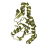
| Title | Crystal structure of the tellurite resistance protein TehB. Northeast Structural Genomics Consortium target VfR98 . | ||||||
 Components Components | Tellurite resistance protein B | ||||||
 Keywords Keywords |  structural genomics / unknown function / X-Ray NESG VfR98 Q5E3X2_VIBF1 / PSI-2 / Protein Structure Initiative / Northeast Structural Genomics Consortium structural genomics / unknown function / X-Ray NESG VfR98 Q5E3X2_VIBF1 / PSI-2 / Protein Structure Initiative / Northeast Structural Genomics Consortium | ||||||
| Function / homology | Tellurite resistance predicted, YeaR / Domain of unknown function DUF1971 / Domain of unknown function (DUF1971) / Jelly Rolls / RmlC-like jelly roll fold / Jelly Rolls / Sandwich / Mainly Beta / Tellurite resistance protein B Function and homology information Function and homology information | ||||||
| Biological species |  Vibrio fischeri ES114 (bacteria) Vibrio fischeri ES114 (bacteria) | ||||||
| Method | X-RAY DIFFRACTION / SYNCHROTRON / SAD / Resolution: 2.3 Å | ||||||
 Authors Authors | Kuzin, A.P. / Su, M. / Seetharaman, J. / Wang, D. / Mao, L. / Maglaqui, M. / Xiao, R. / Liu, J. / Baran, M.C. / Acton, T.B. ...Kuzin, A.P. / Su, M. / Seetharaman, J. / Wang, D. / Mao, L. / Maglaqui, M. / Xiao, R. / Liu, J. / Baran, M.C. / Acton, T.B. / Rost, B. / Montelione, G.T. / Tong, L. / Hunt, J.F. / Northeast Structural Genomics Consortium (NESG) | ||||||
 Citation Citation | Journal: To be Published Title: Crystal structure of the tellurite resistance protein TehB. Northeast Structural Genomics Consortium target VfR98. Authors: Kuzin, A.P. / Su, M. / Seetharaman, J. / Wang, D. / Mao, L. / Maglaqui, M. / Xiao, R. / Liu, J. / Baran, M.C. / Acton, T.B. / Rost, B. / Montelione, G.T. / Tong, L. / Hunt, J.F. | ||||||
| History |
|
- Structure visualization
| Structure viewer | Molecule: MolmilJmol/JSmol |
|---|
- Downloads & links
-Download
| PDBx/mmCIF format | 3dl3.cif.gz | 166.7 KB | Display | PDBx/mmCIF format |
|---|---|---|---|---|
| PDB format | pdb3dl3.ent.gz | 139.1 KB | Display | PDB format |
| PDBx/mmJSON format | 3dl3.json.gz | Tree view | PDBx/mmJSON format | |
| Others |  Other downloads Other downloads |
-Validation report
| Arichive directory | https://data.pdbj.org/pub/pdb/validation_reports/dl/3dl3ftp://data.pdbj.org/pub/pdb/validation_reports/dl/3dl3 | HTTPS FTP |
|---|
-Related structure data
| Similar structure data | |
|---|---|
| Other databases |


-Links
 PDBj
PDBj- Assembly
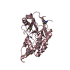
| Deposited unit | 
| ||||||||
|---|---|---|---|---|---|---|---|---|---|
| 1 | 
| ||||||||
| 2 | 
| ||||||||
| 3 | 
| ||||||||
| 4 | 
| ||||||||
| Unit cell |
|
-Components
| #1: Protein | Mass: 13950.108 Da / Num. of mol.: 8 Source method: isolated from a genetically manipulated source Source: (gene. exp.) Vibrio fischeri ES114 (bacteria) / Gene: tehB, VF_1779 / References: UniProt: Q5E3X2#2: Water | ChemComp-HOH / | Water Mass: 18.015 Da / Num. of mol.: 84 / Source method: isolated from a natural source / Formula: H2O Mass: 18.015 Da / Num. of mol.: 84 / Source method: isolated from a natural source / Formula: H2O |
|---|
-Experimental details
-Experiment
| Experiment | Method: X-RAY DIFFRACTION / Number of used crystals: 1 |
|---|
- Sample preparation
| Crystal | Density Matthews: 2.12 Å3/Da / Density % sol: 41.89 % |
|---|---|
| Crystal grow | Temperature: 293 K / pH: 6 Details: 0.1M MgCl2 0.1M MES 40% PEG400, pH 6.0, Crystals were grown by microbatch under oil method. temperature 293K |
-Data collection
| Diffraction |
| |||||||||||||||
|---|---|---|---|---|---|---|---|---|---|---|---|---|---|---|---|---|
| Diffraction source |
| |||||||||||||||
| Detector | Type: ADSC QUANTUM 4 / Detector: CCD / Date: Jun 17, 2008 / Details: mirrors | |||||||||||||||
| Radiation | Protocol: SINGLE WAVELENGTH / Monochromatic (M) / Laue (L): M / Scattering type: x-ray | |||||||||||||||
| Radiation wavelength | Wavelength: 0.979 Å / Relative weight: 1 | |||||||||||||||
| Reflection | Resolution: 2.3→50 Å / Num. obs: 82534 / % possible obs: 97.3 % / Observed criterion σ(I): -3 / Redundancy: 10.8 % / Biso Wilson estimate: 34.6 Å2 / Rmerge(I) obs: 0.072 / Net I/σ(I): 19.6 | |||||||||||||||
| Reflection shell | Resolution: 2.27→2.35 Å / Mean I/σ(I) obs: 3.1 / Rsym value: 0.293 |

- Processing
| Software |
| ||||||||||||||||||||
|---|---|---|---|---|---|---|---|---|---|---|---|---|---|---|---|---|---|---|---|---|---|
| Refinement | Method to determine structure: SAD / Resolution: 2.3→19.9 Å / Rfactor Rfree error: 0.005 / Data cutoff high absF: 320301.99 / Data cutoff low absF: 0 / Isotropic thermal model: RESTRAINED / Cross valid method: THROUGHOUT / σ(F): 1 / Stereochemistry target values: Engh & Huber / Details: BULK SOLVENT MODEL USED
| ||||||||||||||||||||
| Solvent computation | Solvent model: FLAT MODEL / Bsol: 54.9961 Å2 / ksol: 0.45 e/Å3 | ||||||||||||||||||||
| Displacement parameters | Biso mean: 47.7 Å2
| ||||||||||||||||||||
| Refine analyze |
| ||||||||||||||||||||
| Refinement step | Cycle: LAST / Resolution: 2.3→19.9 Å
| ||||||||||||||||||||
| Refine LS restraints |
| ||||||||||||||||||||
| LS refinement shell | Resolution: 2.3→2.44 Å / Rfactor Rfree error: 0.014 / Total num. of bins used: 6
| ||||||||||||||||||||
| Xplor file |
|
