 Movie
Movie Controller


+ Open data

- Basic information
| Entry | Database: PDB / ID: 3dit | ||||||
|---|---|---|---|---|---|---|---|
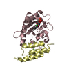
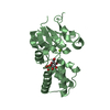
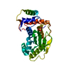
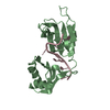
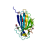
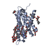
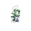
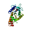
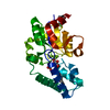
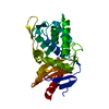
| Title | Crystal structure of MAD MH2 domain | ||||||
 Components Components | Protein mothers against dpp | ||||||
 Keywords Keywords |  SIGNALING PROTEIN / MAD / TGF-beta / MH2 / Cytoplasm / Developmental protein / Nucleus / Phosphoprotein / Transcription / Transcription regulation / Ubl conjugation SIGNALING PROTEIN / MAD / TGF-beta / MH2 / Cytoplasm / Developmental protein / Nucleus / Phosphoprotein / Transcription / Transcription regulation / Ubl conjugation | ||||||
| Function / homology |  Function and homology information Function and homology informationSignaling by BMP / RUNX2 regulates bone development / imaginal disc morphogenesis / R8 cell fate specification / histoblast morphogenesis / negative regulation of salivary gland boundary specification / imaginal disc-derived wing vein morphogenesis / trunk segmentation / imaginal disc-derived leg morphogenesis / germ-line stem cell division ...Signaling by BMP / RUNX2 regulates bone development / imaginal disc morphogenesis / R8 cell fate specification / histoblast morphogenesis / negative regulation of salivary gland boundary specification / imaginal disc-derived wing vein morphogenesis / trunk segmentation / imaginal disc-derived leg morphogenesis / germ-line stem cell division / compound eye morphogenesis / positive regulation of neuromuscular junction development / follicle cell of egg chamber development / wing disc anterior/posterior pattern formation / positive regulation of synaptic assembly at neuromuscular junction / Ub-specific processing proteases / RNA polymerase II transcription repressor complex / intestinal stem cell homeostasis / ventral cord development / co-SMAD binding / heteromeric SMAD protein complex / imaginal disc-derived wing morphogenesis / germ-line stem cell population maintenance / SMAD protein signal transduction / I-SMAD binding / negative regulation of G1/S transition of mitotic cell cycle / regulation of cell differentiation / somatic stem cell population maintenance / anatomical structure morphogenesis / BMP signaling pathway / RNA polymerase II transcription regulatory region sequence-specific DNA binding / DNA-binding transcription repressor activity, RNA polymerase II-specific / heart development / DNA-binding transcription activator activity, RNA polymerase II-specific / transcription regulator complex / RNA polymerase II-specific DNA-binding transcription factor binding / sequence-specific DNA binding / cell differentiation / transcription coactivator activity / DNA-binding transcription factor activity, RNA polymerase II-specific / RNA polymerase II cis-regulatory region sequence-specific DNA binding / regulation of transcription by RNA polymerase II / negative regulation of transcription by RNA polymerase II / positive regulation of transcription by RNA polymerase II / metal ion binding / nucleus / cytosol / cytoplasmSimilarity search - Function | ||||||
| Biological species |  Drosophila melanogaster (fruit fly) Drosophila melanogaster (fruit fly) | ||||||
| Method | X-RAY DIFFRACTION / MOLECULAR REPLACEMENT / molecular replacement / Resolution: 3.2 Å | ||||||
 Authors Authors | Hao, R. / Wu, J.W. / Wang, Z.X. | ||||||
 Citation Citation | Journal: Acta Crystallogr.,Sect.F / Year: 2008 Title: Structure of Drosophila Mad MH2 domain. Authors: Hao, R. / Chen, L. / Wu, J.W. / Wang, Z.X. | ||||||
| History |
|
- Structure visualization
| Structure viewer | Molecule: MolmilJmol/JSmol |
|---|
- Downloads & links
-Download
| PDBx/mmCIF format | 3dit.cif.gz | 122.5 KB | Display | PDBx/mmCIF format |
|---|---|---|---|---|
| PDB format | pdb3dit.ent.gz | 97.3 KB | Display | PDB format |
| PDBx/mmJSON format | 3dit.json.gz | Tree view | PDBx/mmJSON format | |
| Others |  Other downloads Other downloads |
-Validation report
| Arichive directory | https://data.pdbj.org/pub/pdb/validation_reports/di/3ditftp://data.pdbj.org/pub/pdb/validation_reports/di/3dit | HTTPS FTP |
|---|
-Related structure data
| Related structure data | 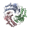 1khuS S: Starting model for refinement |
|---|---|
| Similar structure data |
-Links
 PDBj
PDBj

- Assembly
| Deposited unit | 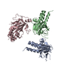
| ||||||||
|---|---|---|---|---|---|---|---|---|---|
| 1 | 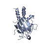
| ||||||||
| 2 | 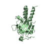
| ||||||||
| 3 | 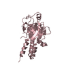
| ||||||||
| Unit cell |
|
-Components
| #1: Protein | Mass: 21312.010 Da / Num. of mol.: 3 / Fragment: MAD MH2 domain Source method: isolated from a genetically manipulated source Source: (gene. exp.) Drosophila melanogaster (fruit fly) / Gene: Mad, CG12399 / Plasmid: pET21b / Production host:  Escherichia coli (E. coli) / Strain (production host): BL21 / References: UniProt: P42003 Escherichia coli (E. coli) / Strain (production host): BL21 / References: UniProt: P42003#2: Water | ChemComp-HOH / | Water Mass: 18.015 Da / Num. of mol.: 90 / Source method: isolated from a natural source / Formula: H2O Mass: 18.015 Da / Num. of mol.: 90 / Source method: isolated from a natural source / Formula: H2O |
|---|
-Experimental details
-Experiment
| Experiment | Method: X-RAY DIFFRACTION / Number of used crystals: 1 |
|---|
- Sample preparation
| Crystal | Density Matthews: 2.77 Å3/Da / Density % sol: 55.57 % |
|---|---|
| Crystal grow | Temperature: 299 K / Method: vapor diffusion, hanging drop / pH: 7.5 Details: 0.1M Hepes, 0.35M Na Formate, 2% Glycerol, pH 7.5, VAPOR DIFFUSION, HANGING DROP, temperature 299K |
-Data collection
| Diffraction | Mean temperature: 100 K |
|---|---|
| Diffraction source | Source: ROTATING ANODE / Type: RIGAKU MICROMAX-007 / Wavelength: 1.5418 Å |
| Detector | Type: RIGAKU RAXIS IV++ / Detector: IMAGE PLATE / Date: Jun 30, 2006 |
| Radiation | Protocol: SINGLE WAVELENGTH / Monochromatic (M) / Laue (L): M / Scattering type: x-ray |
| Radiation wavelength | Wavelength: 1.5418 Å / Relative weight: 1 |
| Reflection | Resolution: 3.2→50 Å / Num. all: 10156 / Num. obs: 10156 / % possible obs: 91.3 % / Observed criterion σ(F): 0 / Observed criterion σ(I): 0 / Redundancy: 7 % / Biso Wilson estimate: 51.6 Å2 / Rmerge(I) obs: 0.116 |
| Reflection shell | Resolution: 3.2→3.31 Å / Redundancy: 7.1 % / Rmerge(I) obs: 0.575 / Mean I/σ(I) obs: 4.01 / Num. unique all: 1126 / % possible all: 100 |
-Phasing
| Phasing | Method: molecular replacement |
|---|
- Processing
| Software |
| ||||||||||||||||||||||||||||
|---|---|---|---|---|---|---|---|---|---|---|---|---|---|---|---|---|---|---|---|---|---|---|---|---|---|---|---|---|---|
| Refinement | Method to determine structure: MOLECULAR REPLACEMENT Starting model: 1KHU Resolution: 3.2→50 Å / σ(F): 0 / σ(I): 0 / Stereochemistry target values: Engh & Huber
| ||||||||||||||||||||||||||||
| Solvent computation | Bsol: 29.977 Å2 | ||||||||||||||||||||||||||||
| Displacement parameters | Biso mean: 63.521 Å2
| ||||||||||||||||||||||||||||
| Refinement step | Cycle: LAST / Resolution: 3.2→50 Å
| ||||||||||||||||||||||||||||
| Refine LS restraints |
| ||||||||||||||||||||||||||||
| Xplor file |
|
