 Movie
Movie Controller

[English] 日本語
 Yorodumi
- PDB-3db9: Crystal structure of UPF0317 protein Atu3911 from Agrobacterium t... -
+ Open data

- Basic information
| Entry | Database: PDB / ID: 3db9 | ||||||
|---|---|---|---|---|---|---|---|
| Title | Crystal structure of UPF0317 protein Atu3911 from Agrobacterium tumefaciens. NorthEast Strcutural Genomics target AtR186 | ||||||
 Components Components | UPF0317 protein Atu3911 | ||||||
 Keywords Keywords |  structural genomics / unknown function / Belongs to the UPF0317 family / Agrobacterium tumefaciens / PSI-2 / Protein Structure Initiative / Northeast Structural Genomics Consortium / NESG structural genomics / unknown function / Belongs to the UPF0317 family / Agrobacterium tumefaciens / PSI-2 / Protein Structure Initiative / Northeast Structural Genomics Consortium / NESG | ||||||
| Function / homology |  Function and homology information Function and homology information | ||||||
| Biological species |  Agrobacterium tumefaciens str. C58 (bacteria) Agrobacterium tumefaciens str. C58 (bacteria) | ||||||
| Method | X-RAY DIFFRACTION / SYNCHROTRON / MOLECULAR REPLACEMENT / Resolution: 2.8 Å | ||||||
 Authors Authors | Seetharaman, J. / Abashidze, M. / Wang, D. / Janjua, H. / Owens, L. / Xiao, R. / Liu, J. / Baran, M.C. / Acton, T.B. / Rost, B. ...Seetharaman, J. / Abashidze, M. / Wang, D. / Janjua, H. / Owens, L. / Xiao, R. / Liu, J. / Baran, M.C. / Acton, T.B. / Rost, B. / Montelione, G.T. / Hunt, J.F. / Tong, L. / Northeast Structural Genomics Consortium (NESG) | ||||||
 Citation Citation | Journal: TO BE PUBLISHED Title: Crystal structure of UPF0317 protein Atu3911 from Agrobacterium tumefaciens. NorthEast Strcutural Genomics target AtR186 (CASP Target) Authors: Seetharaman, J. / Abashidze, M. / Wang, D. / Janjua, H. / Owens, L. / Xiao, R. / Liu, J. / Baran, M.C. / Acton, T.B. / Rost, B. / Montelione, G.T. / Hunt, J.F. / Tong, L. | ||||||
| History |
|
- Structure visualization
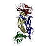
| Structure viewer | Molecule: MolmilJmol/JSmol |
|---|
- Downloads & links
-Download
| PDBx/mmCIF format | 3db9.cif.gz | 59.8 KB | Display | PDBx/mmCIF format |
|---|---|---|---|---|
| PDB format | pdb3db9.ent.gz | 43.1 KB | Display | PDB format |
| PDBx/mmJSON format | 3db9.json.gz | Tree view | PDBx/mmJSON format | |
| Others |  Other downloads Other downloads |
-Validation report
| Arichive directory | https://data.pdbj.org/pub/pdb/validation_reports/db/3db9ftp://data.pdbj.org/pub/pdb/validation_reports/db/3db9 | HTTPS FTP |
|---|
-Related structure data
| Related structure data |  2pifS S: Starting model for refinement |
|---|---|
| Similar structure data | |
| Other databases |


-Links
 PDBj
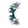
PDBj- Assembly
| Deposited unit | 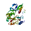
| ||||||||
|---|---|---|---|---|---|---|---|---|---|
| 1 | 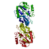
| ||||||||
| 2 |
| ||||||||
| Unit cell |
|
-Components
| #1: Protein | Mass: 28967.020 Da / Num. of mol.: 1 Source method: isolated from a genetically manipulated source Source: (gene. exp.) Agrobacterium tumefaciens str. C58 (bacteria)Strain: Agrobacterium tumefaciens (strain C58 / ATCC 33970 / Gene: Atu3911, AGR_L_1868 / Plasmid: PET21 / Production host: Escherichia coli (E. coli) / References: UniProt: Q8U919 |
|---|---|
| #2: Water | ChemComp-HOH / Water Mass: 18.015 Da / Num. of mol.: 128 / Source method: isolated from a natural source / Formula: H2O Mass: 18.015 Da / Num. of mol.: 128 / Source method: isolated from a natural source / Formula: H2O |
-Experimental details
-Experiment
| Experiment | Method: X-RAY DIFFRACTION / Number of used crystals: 1 |
|---|
- Sample preparation
| Crystal | Density Matthews: 2.16 Å3/Da / Density % sol: 43.03 % |
|---|---|
| Crystal grow | Temperature: 293 K / Method: vapor diffusion, sitting drop / pH: 7 Details: 200 mM NH4SO4, 20% PEG 4000, 50 mM MOPS pH 7. VAPOR DIFFUSION, SITTING DROP, temperature 293K |
-Data collection
| Diffraction | Mean temperature: 100 K |
|---|---|
| Diffraction source | Source: SYNCHROTRON / Site: NSLS  / Beamline: X4A / Wavelength: 0.979 Å / Beamline: X4A / Wavelength: 0.979 Å |
| Detector | Type: ADSC QUANTUM 210 / Detector: CCD / Date: May 2, 2008 / Details: Mirrors |
| Radiation | Protocol: SINGLE WAVELENGTH / Monochromatic (M) / Laue (L): M / Scattering type: x-ray |
| Radiation wavelength | Wavelength: 0.979 Å / Relative weight: 1 |
| Reflection | Resolution: 2.8→50 Å / Num. obs: 11899 / % possible obs: 99.3 % / Observed criterion σ(F): 0 / Observed criterion σ(I): 0 / Redundancy: 21.8 % / Biso Wilson estimate: 105.8 Å2 / Rmerge(I) obs: 0.07 / Rsym value: 0.062 / Net I/σ(I): 17.9 |
| Reflection shell | Resolution: 2.8→2.9 Å / Redundancy: 21 % / Rmerge(I) obs: 0.333 / Mean I/σ(I) obs: 16 / Num. unique all: 1183 / Rsym value: 0.361 / % possible all: 99.6 |
- Processing
| Software |
| ||||||||||||||||||||||||||||||||||||
|---|---|---|---|---|---|---|---|---|---|---|---|---|---|---|---|---|---|---|---|---|---|---|---|---|---|---|---|---|---|---|---|---|---|---|---|---|---|
| Refinement | Method to determine structure: MOLECULAR REPLACEMENT Starting model: 2PIF.pdb Resolution: 2.8→38.94 Å / Rfactor Rfree error: 0.008 / Data cutoff high absF: 226034.91 / Data cutoff low absF: 0 / Isotropic thermal model: RESTRAINED / Cross valid method: THROUGHOUT / σ(F): 2 / Stereochemistry target values: Engh & Huber
| ||||||||||||||||||||||||||||||||||||
| Solvent computation | Solvent model: FLAT MODEL / Bsol: 67.4671 Å2 / ksol: 0.368438 e/Å3 | ||||||||||||||||||||||||||||||||||||
| Displacement parameters | Biso mean: 54.4 Å2
| ||||||||||||||||||||||||||||||||||||
| Refine analyze |
| ||||||||||||||||||||||||||||||||||||
| Refinement step | Cycle: LAST / Resolution: 2.8→38.94 Å
| ||||||||||||||||||||||||||||||||||||
| Refine LS restraints |
| ||||||||||||||||||||||||||||||||||||
| LS refinement shell | Resolution: 2.8→2.98 Å / Rfactor Rfree error: 0.031 / Total num. of bins used: 6
| ||||||||||||||||||||||||||||||||||||
| Xplor file |
|
