 Movie
Movie Controller

[English] 日本語
 Yorodumi
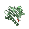
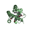
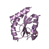
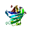
- PDB-3dav: Schizosaccharomyces Pombe Profilin crystallized from Sodium formate -
+ Open data

- Basic information
| Entry | Database: PDB / ID: 3dav | ||||||
|---|---|---|---|---|---|---|---|
| Title | Schizosaccharomyces Pombe Profilin crystallized from Sodium formate | ||||||
 Components Components | Profilin | ||||||
 Keywords Keywords | PROTEIN BINDING / profilin / yeast / pombe / protein-protein interaction / Actin-binding / Cytoplasm / Cytoskeleton | ||||||
| Function / homology |  Function and homology information Function and homology informationcytogamy / actin cortical patch organization / cell cortex of cell tip / medial cortex / mitotic actomyosin contractile ring assembly / actin cortical patch / sequestering of actin monomers / mating projection tip / actin monomer binding / actin filament polymerization ...cytogamy / actin cortical patch organization / cell cortex of cell tip / medial cortex / mitotic actomyosin contractile ring assembly / actin cortical patch / sequestering of actin monomers / mating projection tip / actin monomer binding / actin filament polymerization / guanyl-nucleotide exchange factor activity / cell cortexSimilarity search - Function | ||||||
| Biological species |  Schizosaccharomyces pombe (fission yeast) Schizosaccharomyces pombe (fission yeast) | ||||||
| Method | X-RAY DIFFRACTION / Resolution: 2.2 Å | ||||||
 Authors Authors | Ezezika, O.C. / Nolen, B.J. / Pollard, T.D. | ||||||
 Citation Citation | Journal: J.Biol.Chem. / Year: 2009 Title: Incompatibility with Formin Cdc12p Prevents Human Profilin from Substituting for Fission Yeast Profilin: INSIGHTS FROM CRYSTAL STRUCTURES OF FISSION YEAST PROFILIN. Authors: Ezezika, O.C. / Younger, N.S. / Lu, J. / Kaiser, D.A. / Corbin, Z.A. / Nolen, B.J. / Kovar, D.R. / Pollard, T.D. | ||||||
| History |
|
- Structure visualization
| Structure viewer | Molecule: MolmilJmol/JSmol |
|---|
- Downloads & links
-Download
| PDBx/mmCIF format | 3dav.cif.gz | 64.2 KB | Display | PDBx/mmCIF format |
|---|---|---|---|---|
| PDB format | pdb3dav.ent.gz | 46.5 KB | Display | PDB format |
| PDBx/mmJSON format | 3dav.json.gz | Tree view | PDBx/mmJSON format | |
| Others |  Other downloads Other downloads |
-Validation report
| Arichive directory | https://data.pdbj.org/pub/pdb/validation_reports/da/3davftp://data.pdbj.org/pub/pdb/validation_reports/da/3dav | HTTPS FTP |
|---|
-Related structure data
| Related structure data | 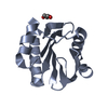 3d9ySC S: Starting model for refinement C: citing same article ( |
|---|---|
| Similar structure data |


-Links
 PDBj
PDBj
- Assembly
| Deposited unit | 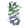
| ||||||||
|---|---|---|---|---|---|---|---|---|---|
| 1 | 
| ||||||||
| 2 | 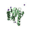
| ||||||||
| Unit cell |
|
-Components
| #1: Protein | Mass: 13422.347 Da / Num. of mol.: 2 Source method: isolated from a genetically manipulated source Source: (gene. exp.) Schizosaccharomyces pombe (fission yeast)Strain: Schizosaccharomyces pombe / Gene: cdc3, SPAC4A8.15c / Plasmid: pMW-SpPRF / Production host:  Escherichia coli (E. coli) / Strain (production host): B21(DE3) / References: UniProt: P39825 Escherichia coli (E. coli) / Strain (production host): B21(DE3) / References: UniProt: P39825#2: Chemical | ChemComp-NA /   Mass: 22.990 Da / Num. of mol.: 9 / Source method: obtained synthetically / Formula: Na Mass: 22.990 Da / Num. of mol.: 9 / Source method: obtained synthetically / Formula: Na#3: Water | ChemComp-HOH / | Water Mass: 18.015 Da / Num. of mol.: 231 / Source method: isolated from a natural source / Formula: H2O Mass: 18.015 Da / Num. of mol.: 231 / Source method: isolated from a natural source / Formula: H2O |
|---|
-Experimental details
-Experiment
| Experiment | Method: X-RAY DIFFRACTION / Number of used crystals: 1 |
|---|
- Sample preparation
| Crystal | Density Matthews: 2.19 Å3/Da / Density % sol: 43.75 % |
|---|---|
| Crystal grow | Temperature: 296.15 K / Method: vapor diffusion, hanging drop Details: 4.0 M sodium formate , VAPOR DIFFUSION, HANGING DROP, temperature 296.15K |
-Data collection
| Diffraction | Mean temperature: 273 K |
|---|---|
| Diffraction source | Source: ROTATING ANODE / Type: OTHER / Wavelength: 1.54 Å |
| Detector | Type: MAR scanner 345 mm plate / Detector: IMAGE PLATE / Date: Mar 17, 2008 |
| Radiation | Monochromator: Mirrors / Protocol: SINGLE WAVELENGTH / Monochromatic (M) / Laue (L): M / Scattering type: x-ray |
| Radiation wavelength | Wavelength: 1.54 Å / Relative weight: 1 |
| Reflection | Resolution: 2.2→50 Å / Num. obs: 11429 / % possible obs: 95.8 % / Redundancy: 2.9 % / Rmerge(I) obs: 0.09 / Rsym value: 0.069 / Net I/σ(I): 15.28 |
| Reflection shell | Resolution: 2.2→2.27 Å / Redundancy: 2.2 % / Rmerge(I) obs: 0.318 / Mean I/σ(I) obs: 4.03 / Num. unique all: 747 / Rsym value: 0.343 / % possible all: 64 |
- Processing
| Software |
| |||||||||||||||||||||||||||||||||||||||||||||||||||||||||||||||||||||||||||||||||||||||||||||||
|---|---|---|---|---|---|---|---|---|---|---|---|---|---|---|---|---|---|---|---|---|---|---|---|---|---|---|---|---|---|---|---|---|---|---|---|---|---|---|---|---|---|---|---|---|---|---|---|---|---|---|---|---|---|---|---|---|---|---|---|---|---|---|---|---|---|---|---|---|---|---|---|---|---|---|---|---|---|---|---|---|---|---|---|---|---|---|---|---|---|---|---|---|---|---|---|---|
| Refinement | Starting model: 3D9Y Resolution: 2.2→41.2 Å / Cor.coef. Fo:Fc: 0.944 / Cor.coef. Fo:Fc free: 0.884 / SU B: 12.947 / SU ML: 0.174 / Cross valid method: THROUGHOUT / σ(F): 0 / ESU R: 0.37 / ESU R Free: 0.259 / Stereochemistry target values: MAXIMUM LIKELIHOOD
| |||||||||||||||||||||||||||||||||||||||||||||||||||||||||||||||||||||||||||||||||||||||||||||||
| Solvent computation | Ion probe radii: 0.8 Å / Shrinkage radii: 0.8 Å / VDW probe radii: 1.2 Å / Solvent model: BABINET MODEL WITH MASK | |||||||||||||||||||||||||||||||||||||||||||||||||||||||||||||||||||||||||||||||||||||||||||||||
| Displacement parameters | Biso mean: 24.058 Å2
| |||||||||||||||||||||||||||||||||||||||||||||||||||||||||||||||||||||||||||||||||||||||||||||||
| Refinement step | Cycle: LAST / Resolution: 2.2→41.2 Å
| |||||||||||||||||||||||||||||||||||||||||||||||||||||||||||||||||||||||||||||||||||||||||||||||
| Refine LS restraints |
| |||||||||||||||||||||||||||||||||||||||||||||||||||||||||||||||||||||||||||||||||||||||||||||||
| LS refinement shell | Resolution: 2.2→2.258 Å / Total num. of bins used: 20
|
