 ムービー
ムービー コントローラー


+ データを開く

- 基本情報
基本情報
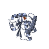
| 登録情報 | データベース: PDB / ID: 3d8e | ||||||
|---|---|---|---|---|---|---|---|
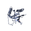
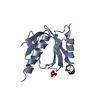
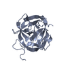
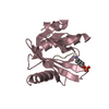
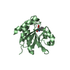
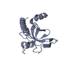
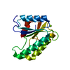
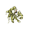
| タイトル | Crystal structure of the human Fe65-PTB1 domain (trigonal crystal form) | ||||||
 要素 要素 | Amyloid beta A4 precursor protein-binding family B member 1 | ||||||
 キーワード キーワード |  PROTEIN BINDING (タンパク質) / alpha-beta structure / phosphotyrosine binding domain PROTEIN BINDING (タンパク質) / alpha-beta structure / phosphotyrosine binding domain | ||||||
| 機能・相同性 |  機能・相同性情報 機能・相同性情報negative regulation of cell cycle G1/S phase transition / proline-rich region binding / low-density lipoprotein particle receptor binding / smooth muscle contraction / 軸索誘導 / positive regulation of protein secretion / positive regulation of neuron projection development / Recruitment and ATM-mediated phosphorylation of repair and signaling proteins at DNA double strand breaks / lamellipodium / chromatin organization ...negative regulation of cell cycle G1/S phase transition / proline-rich region binding / low-density lipoprotein particle receptor binding / smooth muscle contraction / 軸索誘導 / positive regulation of protein secretion / positive regulation of neuron projection development / Recruitment and ATM-mediated phosphorylation of repair and signaling proteins at DNA double strand breaks / lamellipodium / chromatin organization / amyloid-beta binding / histone binding / 成長円錐 / transcription coactivator activity / molecular adaptor activity / nuclear speck / positive regulation of apoptotic process / apoptotic process / シナプス / DNA damage response / ubiquitin protein ligase binding / chromatin binding / regulation of DNA-templated transcription / positive regulation of DNA-templated transcription / negative regulation of transcription by RNA polymerase II / 小胞体 / シグナル伝達 / positive regulation of transcription by RNA polymerase II / 核質 / 細胞核 / 細胞膜 / 細胞質類似検索 - 分子機能 | ||||||
| 生物種 |  Homo sapiens (ヒト) Homo sapiens (ヒト) | ||||||
| 手法 | X線回折 / シンクロトロン / 解像度: 2.8 Å | ||||||
 データ登録者 データ登録者 | Radzimanowski, J. / Ravaud, S. / Sinning, I. / Wild, K. | ||||||
 引用 引用 | ジャーナル: J.Biol.Chem. / 年: 2008 タイトル: Crystal structure of the human Fe65-PTB1 domain. 著者: Radzimanowski, J. / Ravaud, S. / Schlesinger, S. / Koch, J. / Beyreuther, K. / Sinning, I. / Wild, K. #1: ジャーナル: Acta Crystallogr.,Sect.F / 年: 2008 タイトル: Mercury-induced crystallization and SAD phasing of the human Fe65-PTB1 domain. 著者: Radzimanowski, J. / Ravaud, S. / Beyreuther, K. / Sinning, I. / Wild, K. | ||||||
| 履歴 |
|
- 構造の表示
構造の表示
| 構造ビューア | 分子: MolmilJmol/JSmol |
|---|
- ダウンロードとリンク
-ダウンロード
| PDBx/mmCIF形式 | 3d8e.cif.gz | 104.9 KB | 表示 | PDBx/mmCIF形式 |
|---|---|---|---|---|
| PDB形式 | pdb3d8e.ent.gz | 82.7 KB | 表示 | PDB形式 |
| PDBx/mmJSON形式 | 3d8e.json.gz | ツリー表示 | PDBx/mmJSON形式 | |
| その他 |  その他のダウンロード その他のダウンロード |
-検証レポート
| アーカイブディレクトリ | https://data.pdbj.org/pub/pdb/validation_reports/d8/3d8eftp://data.pdbj.org/pub/pdb/validation_reports/d8/3d8e | HTTPS FTP |
|---|
-関連構造データ


-リンク
 PDBj
PDBj

- 集合体
集合体
| 登録構造単位 | 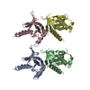
| ||||||||
|---|---|---|---|---|---|---|---|---|---|
| 1 | 
| ||||||||
| 2 | 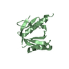
| ||||||||
| 3 | 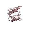
| ||||||||
| 4 | 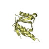
| ||||||||
| 単位格子 |
|
-要素
| #1: タンパク質 | 分子量: 16782.211 Da / 分子数: 4 / 断片: Phosphotyrosine binding domain 1 / 由来タイプ: 組換発現 / 由来: (組換発現) Homo sapiens (ヒト) / 遺伝子: APBB1, FE65, RIR / プラスミド: pET24d / 発現宿主:  Escherichia coli (大腸菌) / 株 (発現宿主): BL21 (DE3) / 参照: UniProt: O00213 Escherichia coli (大腸菌) / 株 (発現宿主): BL21 (DE3) / 参照: UniProt: O00213 |
|---|
-実験情報
-実験
| 実験 | 手法: X線回折 / 使用した結晶の数: 1 |
|---|
- 試料調製
試料調製
| 結晶 | マシュー密度: 2.41 Å3/Da / 溶媒含有率: 49 % |
|---|---|
| 結晶化 | 温度: 291 K / 手法: 蒸気拡散法 / pH: 7.5 詳細: 100mM HEPES, 5% (v/v) ethylene glycol, 10% (w/v) PEG 3350, pH 7.5, VAPOR DIFFUSION, temperature 291K |
-データ収集
| 回折 | 平均測定温度: 100 K |
|---|---|
| 放射光源 | 由来: シンクロトロン / サイト: ESRF  / ビームライン: ID14-2 / 波長: 1.0723 Å / ビームライン: ID14-2 / 波長: 1.0723 Å |
| 検出器 | タイプ: ADSC QUANTUM 4 / 検出器: CCD / 日付: 2007年7月19日 |
| 放射 | プロトコル: SINGLE WAVELENGTH / 単色(M)・ラウエ(L): M / 散乱光タイプ: x-ray |
| 放射波長 | 波長: 1.0723 Å / 相対比: 1 |
| 反射 | 解像度: 2.8→72.9 Å / Num. obs: 15431 / % possible obs: 99.8 % / Observed criterion σ(F): 0 / Observed criterion σ(I): 0 / 冗長度: 3.6 % / Rmerge(I) obs: 0.089 / Net I/σ(I): 14.2 |
| 反射 シェル | 解像度: 2.8→2.95 Å / 冗長度: 3.7 % / Rmerge(I) obs: 0.347 / Mean I/σ(I) obs: 3.2 / % possible all: 99.9 |
- 解析
解析
| ソフトウェア |
| ||||||||||||||||||||||||||||||||||||||||||||||||||||||||||||||||||||||||||||||||||||||||||
|---|---|---|---|---|---|---|---|---|---|---|---|---|---|---|---|---|---|---|---|---|---|---|---|---|---|---|---|---|---|---|---|---|---|---|---|---|---|---|---|---|---|---|---|---|---|---|---|---|---|---|---|---|---|---|---|---|---|---|---|---|---|---|---|---|---|---|---|---|---|---|---|---|---|---|---|---|---|---|---|---|---|---|---|---|---|---|---|---|---|---|---|
| 精密化 | 解像度: 2.8→25 Å / Cor.coef. Fo:Fc: 0.927 / Cor.coef. Fo:Fc free: 0.881 / SU B: 16.209 / SU ML: 0.329 / 交差検証法: THROUGHOUT / σ(F): 0 / ESU R Free: 0.463 / 立体化学のターゲット値: MAXIMUM LIKELIHOOD
| ||||||||||||||||||||||||||||||||||||||||||||||||||||||||||||||||||||||||||||||||||||||||||
| 溶媒の処理 | イオンプローブ半径: 0.8 Å / 減衰半径: 0.8 Å / VDWプローブ半径: 1.4 Å / 溶媒モデル: MASK | ||||||||||||||||||||||||||||||||||||||||||||||||||||||||||||||||||||||||||||||||||||||||||
| 原子変位パラメータ | Biso mean: 59.775 Å2
| ||||||||||||||||||||||||||||||||||||||||||||||||||||||||||||||||||||||||||||||||||||||||||
| Refine analyze | Luzzati sigma a obs: 0.396 Å | ||||||||||||||||||||||||||||||||||||||||||||||||||||||||||||||||||||||||||||||||||||||||||
| 精密化ステップ | サイクル: LAST / 解像度: 2.8→25 Å
| ||||||||||||||||||||||||||||||||||||||||||||||||||||||||||||||||||||||||||||||||||||||||||
| 拘束条件 |
| ||||||||||||||||||||||||||||||||||||||||||||||||||||||||||||||||||||||||||||||||||||||||||
| LS精密化 シェル | 解像度: 2.8→2.872 Å / Total num. of bins used: 20
|
