 Movie
Movie Controller

[English] 日本語
 Yorodumi
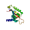
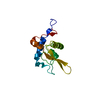
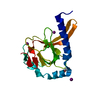
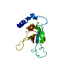
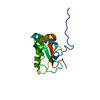
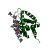
- PDB-3d89: Crystal Structure of a Soluble Rieske Ferredoxin from Mus musculus -
+ Open data

- Basic information
| Entry | Database: PDB / ID: 3d89 | ||||||
|---|---|---|---|---|---|---|---|
| Title | Crystal Structure of a Soluble Rieske Ferredoxin from Mus musculus | ||||||
 Components Components | Rieske domain-containing protein | ||||||
 Keywords Keywords |  ELECTRON TRANSPORT / CASP Target / Rieske ferredoxin / [2Fe-2S] cluster / Protein Structure Initiative / PSI / Center for Eukaryotic Structural Genomics / CESG / Iron / Iron-sulfur / Metal-binding ELECTRON TRANSPORT / CASP Target / Rieske ferredoxin / [2Fe-2S] cluster / Protein Structure Initiative / PSI / Center for Eukaryotic Structural Genomics / CESG / Iron / Iron-sulfur / Metal-binding | ||||||
| Function / homology |  Function and homology information Function and homology information | ||||||
| Biological species |  Mus musculus (house mouse) Mus musculus (house mouse) | ||||||
| Method | X-RAY DIFFRACTION / SYNCHROTRON / SAD / Resolution: 2.071 Å | ||||||
 Authors Authors | Levin, E.J. / McCoy, J.G. / Elsen, N.L. / Seder, K.D. / Bingman, C.A. / Wesenberg, G.E. / Fox, B.G. / Phillips Jr., G.N. / Center for Eukaryotic Structural Genomics (CESG) | ||||||
 Citation Citation | Journal: Acta Crystallogr.,Sect.D / Year: 2008 Title: X-ray structure of a soluble Rieske-type ferredoxin from Mus musculus. Authors: Levin, E.J. / Elsen, N.L. / Seder, K.D. / McCoy, J.G. / Fox, B.G. / Phillips, G.N. | ||||||
| History |
|
- Structure visualization
| Structure viewer | Molecule: MolmilJmol/JSmol |
|---|
- Downloads & links
-Download
| PDBx/mmCIF format | 3d89.cif.gz | 45.9 KB | Display | PDBx/mmCIF format |
|---|---|---|---|---|
| PDB format | pdb3d89.ent.gz | 30.9 KB | Display | PDB format |
| PDBx/mmJSON format | 3d89.json.gz | Tree view | PDBx/mmJSON format | |
| Others |  Other downloads Other downloads |
-Validation report
| Arichive directory | https://data.pdbj.org/pub/pdb/validation_reports/d8/3d89ftp://data.pdbj.org/pub/pdb/validation_reports/d8/3d89 | HTTPS FTP |
|---|
-Related structure data
| Similar structure data | |
|---|---|
| Other databases |


-Links
 PDBj
PDBj


- Assembly
| Deposited unit | 
| ||||||||
|---|---|---|---|---|---|---|---|---|---|
| 1 |
| ||||||||
| Unit cell |
| ||||||||
| Details | authors state that Gel-filtration data has shown that in solution the protein is a monomer and the PISA calculated asymmetric dimer is elieved to be an artifact of the lattice packing. |
-Components
| #1: Protein | Mass: 17951.223 Da / Num. of mol.: 1 Source method: isolated from a genetically manipulated source Source: (gene. exp.) Mus musculus (house mouse) / Gene: Rfesd / Plasmid: PVP 16 / Production host:  Escherichia coli (E. coli) / Strain (production host): BL21 / References: UniProt: Q8K2P6 Escherichia coli (E. coli) / Strain (production host): BL21 / References: UniProt: Q8K2P6 |
|---|---|
| #2: Chemical | ChemComp-FES / Iron–sulfur cluster  Mass: 175.820 Da / Num. of mol.: 1 / Source method: obtained synthetically / Formula: Fe2S2 Mass: 175.820 Da / Num. of mol.: 1 / Source method: obtained synthetically / Formula: Fe2S2 |
| #3: Chemical | ChemComp-EDO / Ethylene glycol  Mass: 62.068 Da / Num. of mol.: 1 / Source method: obtained synthetically / Formula: C2H6O2 Mass: 62.068 Da / Num. of mol.: 1 / Source method: obtained synthetically / Formula: C2H6O2 |
| #4: Water | ChemComp-HOH / Water Mass: 18.015 Da / Num. of mol.: 48 / Source method: isolated from a natural source / Formula: H2O Mass: 18.015 Da / Num. of mol.: 48 / Source method: isolated from a natural source / Formula: H2O |
| Sequence details | THIS STRUCTURE WAS SUBMITTED AS CASP8 ID T0391. |
-Experimental details
-Experiment
| Experiment | Method: X-RAY DIFFRACTION / Number of used crystals: 2 |
|---|
- Sample preparation
| Crystal | Density Matthews: 2.1 Å3/Da / Density % sol: 40.5 % |
|---|---|
| Crystal grow | Temperature: 293 K / Method: vapor diffusion, hanging drop / pH: 6 Details: Protein Solution (~15 mg/ml native protein, 0.025 M NaCl, 0.005 M MOPS pH 7.0) mixed in a 1:1 ratio with Well Solution ( 25% MEPEG 5K, 0.12 M Trisodium citrate, 0.10 M MES ph 6.0) ...Details: Protein Solution (~15 mg/ml native protein, 0.025 M NaCl, 0.005 M MOPS pH 7.0) mixed in a 1:1 ratio with Well Solution ( 25% MEPEG 5K, 0.12 M Trisodium citrate, 0.10 M MES ph 6.0) Cryoprotected with 15% ethylene glycol; Mercury derivative crystal soaked overnight in in mother liquor containing 0.002 M thimersol, vapor diffusion, hanging drop, temperature 293K, VAPOR DIFFUSION, HANGING DROP |
-Data collection
| Diffraction |
| |||||||||||||||||||||||||||||||||||||||||||||||||||||||||||||||||||||||||||||
|---|---|---|---|---|---|---|---|---|---|---|---|---|---|---|---|---|---|---|---|---|---|---|---|---|---|---|---|---|---|---|---|---|---|---|---|---|---|---|---|---|---|---|---|---|---|---|---|---|---|---|---|---|---|---|---|---|---|---|---|---|---|---|---|---|---|---|---|---|---|---|---|---|---|---|---|---|---|---|
| Diffraction source |
| |||||||||||||||||||||||||||||||||||||||||||||||||||||||||||||||||||||||||||||
| Detector |
| |||||||||||||||||||||||||||||||||||||||||||||||||||||||||||||||||||||||||||||
| Radiation |
| |||||||||||||||||||||||||||||||||||||||||||||||||||||||||||||||||||||||||||||
| Radiation wavelength |
| |||||||||||||||||||||||||||||||||||||||||||||||||||||||||||||||||||||||||||||
| Reflection | Resolution: 2.07→50 Å / Num. obs: 9806 / % possible obs: 99.4 % / Redundancy: 12.7 % / Rmerge(I) obs: 0.057 / Χ2: 0.907 / Net I/σ(I): 18.354 | |||||||||||||||||||||||||||||||||||||||||||||||||||||||||||||||||||||||||||||
| Reflection shell | Diffraction-ID: 1,2
|

-Phasing
| Phasing | Method: SAD | ||||||||||||||||||||||||||||||||||||||||||||||||||||||||||||||||||||||||||||||||||||||||||||||||||||||||||||||||||||||||||||||||||||||||||||||||||||||||||||||||||||||||||||||||||||||||||||||||||||||||||||||||||||||||||||||||||||||||||||||||||||||
|---|---|---|---|---|---|---|---|---|---|---|---|---|---|---|---|---|---|---|---|---|---|---|---|---|---|---|---|---|---|---|---|---|---|---|---|---|---|---|---|---|---|---|---|---|---|---|---|---|---|---|---|---|---|---|---|---|---|---|---|---|---|---|---|---|---|---|---|---|---|---|---|---|---|---|---|---|---|---|---|---|---|---|---|---|---|---|---|---|---|---|---|---|---|---|---|---|---|---|---|---|---|---|---|---|---|---|---|---|---|---|---|---|---|---|---|---|---|---|---|---|---|---|---|---|---|---|---|---|---|---|---|---|---|---|---|---|---|---|---|---|---|---|---|---|---|---|---|---|---|---|---|---|---|---|---|---|---|---|---|---|---|---|---|---|---|---|---|---|---|---|---|---|---|---|---|---|---|---|---|---|---|---|---|---|---|---|---|---|---|---|---|---|---|---|---|---|---|---|---|---|---|---|---|---|---|---|---|---|---|---|---|---|---|---|---|---|---|---|---|---|---|---|---|---|---|---|---|---|---|---|---|---|---|---|---|---|---|---|---|---|---|---|---|---|---|---|---|
| Phasing MAD set | R cullis centric: 0 / Highest resolution: 1.69 Å / Lowest resolution: 30.66 Å / Power centric: 0
| ||||||||||||||||||||||||||||||||||||||||||||||||||||||||||||||||||||||||||||||||||||||||||||||||||||||||||||||||||||||||||||||||||||||||||||||||||||||||||||||||||||||||||||||||||||||||||||||||||||||||||||||||||||||||||||||||||||||||||||||||||||||
| Phasing MAD set shell | R cullis centric: 0 / Power centric: 0
| ||||||||||||||||||||||||||||||||||||||||||||||||||||||||||||||||||||||||||||||||||||||||||||||||||||||||||||||||||||||||||||||||||||||||||||||||||||||||||||||||||||||||||||||||||||||||||||||||||||||||||||||||||||||||||||||||||||||||||||||||||||||
| Phasing MAD set site |
| ||||||||||||||||||||||||||||||||||||||||||||||||||||||||||||||||||||||||||||||||||||||||||||||||||||||||||||||||||||||||||||||||||||||||||||||||||||||||||||||||||||||||||||||||||||||||||||||||||||||||||||||||||||||||||||||||||||||||||||||||||||||
| Phasing dm | Method: Solvent flattening and Histogram matching / Reflection: 17514 | ||||||||||||||||||||||||||||||||||||||||||||||||||||||||||||||||||||||||||||||||||||||||||||||||||||||||||||||||||||||||||||||||||||||||||||||||||||||||||||||||||||||||||||||||||||||||||||||||||||||||||||||||||||||||||||||||||||||||||||||||||||||
| Phasing dm shell |
|
- Processing
| Software |
| |||||||||||||||||||||||||||||||||||||||||||||||||||||||||||||||||||||||||||||||||||||||||||||||||||||||||||||||||||||||||||||
|---|---|---|---|---|---|---|---|---|---|---|---|---|---|---|---|---|---|---|---|---|---|---|---|---|---|---|---|---|---|---|---|---|---|---|---|---|---|---|---|---|---|---|---|---|---|---|---|---|---|---|---|---|---|---|---|---|---|---|---|---|---|---|---|---|---|---|---|---|---|---|---|---|---|---|---|---|---|---|---|---|---|---|---|---|---|---|---|---|---|---|---|---|---|---|---|---|---|---|---|---|---|---|---|---|---|---|---|---|---|---|---|---|---|---|---|---|---|---|---|---|---|---|---|---|---|---|
| Refinement | Method to determine structure: SAD / Resolution: 2.071→37.75 Å / Cor.coef. Fo:Fc: 0.959 / Cor.coef. Fo:Fc free: 0.945 / WRfactor Rfree: 0.245 / WRfactor Rwork: 0.218 / SU B: 7.655 / SU ML: 0.124 / TLS residual ADP flag: LIKELY RESIDUAL / Cross valid method: THROUGHOUT / σ(F): 0 / ESU R: 0.213 / ESU R Free: 0.171 / Stereochemistry target values: MAXIMUM LIKELIHOOD / Details: HYDROGENS HAVE BEEN ADDED IN THE RIDING POSITIONS
| |||||||||||||||||||||||||||||||||||||||||||||||||||||||||||||||||||||||||||||||||||||||||||||||||||||||||||||||||||||||||||||
| Solvent computation | Ion probe radii: 0.8 Å / Shrinkage radii: 0.8 Å / VDW probe radii: 1.2 Å / Solvent model: MASK BULK SOLVENT | |||||||||||||||||||||||||||||||||||||||||||||||||||||||||||||||||||||||||||||||||||||||||||||||||||||||||||||||||||||||||||||
| Displacement parameters | Biso mean: 46.027 Å2
| |||||||||||||||||||||||||||||||||||||||||||||||||||||||||||||||||||||||||||||||||||||||||||||||||||||||||||||||||||||||||||||
| Refinement step | Cycle: LAST / Resolution: 2.071→37.75 Å
| |||||||||||||||||||||||||||||||||||||||||||||||||||||||||||||||||||||||||||||||||||||||||||||||||||||||||||||||||||||||||||||
| Refine LS restraints |
| |||||||||||||||||||||||||||||||||||||||||||||||||||||||||||||||||||||||||||||||||||||||||||||||||||||||||||||||||||||||||||||
| LS refinement shell | Resolution: 2.071→2.125 Å / Total num. of bins used: 20
| |||||||||||||||||||||||||||||||||||||||||||||||||||||||||||||||||||||||||||||||||||||||||||||||||||||||||||||||||||||||||||||
| Refinement TLS params. | Method: refined / Refine-ID: X-RAY DIFFRACTION
| |||||||||||||||||||||||||||||||||||||||||||||||||||||||||||||||||||||||||||||||||||||||||||||||||||||||||||||||||||||||||||||
| Refinement TLS group |
|
