 Movie
Movie Controller

[English] 日本語
 Yorodumi
- PDB-3d3h: Crystal structure of a complex of the peptidoglycan glycosyltrans... -
+ Open data

- Basic information
| Entry | Database: PDB / ID: 3d3h | ||||||
|---|---|---|---|---|---|---|---|
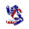
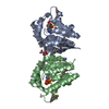
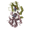
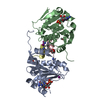
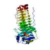
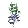
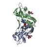
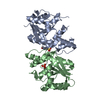
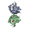
| Title | Crystal structure of a complex of the peptidoglycan glycosyltransferase domain from Aquifex aeolicus and neryl moenomycin A | ||||||
 Components Components | Penicillin-insensitive transglycosylase | ||||||
 Keywords Keywords | TRANSFERASE/ANTIBIOTIC /  peptidoglycan glycosyltransferase / cell wall biosynthesis / antibiotics / penicillin-binding protein / transglycosylase / moenomycin / Antibiotic resistance / Cell shape / Cell wall biogenesis/degradation / Hydrolase / Inner membrane / Membrane / Multifunctional enzyme / Peptidoglycan synthesis / Signal-anchor / Transmembrane / TRANSFERASE-ANTIBIOTIC COMPLEX peptidoglycan glycosyltransferase / cell wall biosynthesis / antibiotics / penicillin-binding protein / transglycosylase / moenomycin / Antibiotic resistance / Cell shape / Cell wall biogenesis/degradation / Hydrolase / Inner membrane / Membrane / Multifunctional enzyme / Peptidoglycan synthesis / Signal-anchor / Transmembrane / TRANSFERASE-ANTIBIOTIC COMPLEX | ||||||
| Function / homology |  Function and homology informationpeptidoglycan glycosyltransferase / peptidoglycan glycosyltransferase activity / serine-type D-Ala-D-Ala carboxypeptidase / serine-type D-Ala-D-Ala carboxypeptidase activity / penicillin binding / peptidoglycan biosynthetic process / cell wall organization / regulation of cell shape / response to antibiotic / proteolysis ...peptidoglycan glycosyltransferase / peptidoglycan glycosyltransferase activity / serine-type D-Ala-D-Ala carboxypeptidase / serine-type D-Ala-D-Ala carboxypeptidase activity / penicillin binding / peptidoglycan biosynthetic process / cell wall organization / regulation of cell shape / response to antibiotic / proteolysis / identical protein binding / plasma membrane Function and homology informationpeptidoglycan glycosyltransferase / peptidoglycan glycosyltransferase activity / serine-type D-Ala-D-Ala carboxypeptidase / serine-type D-Ala-D-Ala carboxypeptidase activity / penicillin binding / peptidoglycan biosynthetic process / cell wall organization / regulation of cell shape / response to antibiotic / proteolysis ...peptidoglycan glycosyltransferase / peptidoglycan glycosyltransferase activity / serine-type D-Ala-D-Ala carboxypeptidase / serine-type D-Ala-D-Ala carboxypeptidase activity / penicillin binding / peptidoglycan biosynthetic process / cell wall organization / regulation of cell shape / response to antibiotic / proteolysis / identical protein binding / plasma membraneSimilarity search - Function | ||||||
| Biological species |   Aquifex aeolicus (bacteria) Aquifex aeolicus (bacteria) | ||||||
| Method | X-RAY DIFFRACTION / SYNCHROTRON / MOLECULAR REPLACEMENT / Resolution: 2.31 Å | ||||||
 Authors Authors | Yuan, Y. / Sliz, P. / Walker, S. | ||||||
 Citation Citation | Journal: Acs Chem.Biol. / Year: 2008 Title: Structural analysis of the contacts anchoring moenomycin to peptidoglycan glycosyltransferases and implications for antibiotic design. Authors: Yuan, Y. / Fuse, S. / Ostash, B. / Sliz, P. / Kahne, D. / Walker, S. | ||||||
| History |
|
- Structure visualization
| Structure viewer | Molecule: MolmilJmol/JSmol |
|---|
- Downloads & links
-Download
| PDBx/mmCIF format | 3d3h.cif.gz | 53.7 KB | Display | PDBx/mmCIF format |
|---|---|---|---|---|
| PDB format | pdb3d3h.ent.gz | 37.3 KB | Display | PDB format |
| PDBx/mmJSON format | 3d3h.json.gz | Tree view | PDBx/mmJSON format | |
| Others |  Other downloads Other downloads |
-Validation report
| Arichive directory | https://data.pdbj.org/pub/pdb/validation_reports/d3/3d3hftp://data.pdbj.org/pub/pdb/validation_reports/d3/3d3h | HTTPS FTP |
|---|
-Related structure data
| Related structure data | 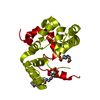 2oqoS S: Starting model for refinement |
|---|---|
| Similar structure data |

-Links
 PDBj
PDBj
- Assembly
| Deposited unit | 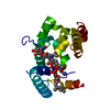
| ||||||||
|---|---|---|---|---|---|---|---|---|---|
| 1 | 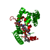
| ||||||||
| Unit cell |
|
-Components
| #1: Protein | Mass: 22912.428 Da / Num. of mol.: 1 Source method: isolated from a genetically manipulated source Source: (gene. exp.) Aquifex aeolicus (bacteria) / Strain: VF5 / Gene: mrcA, ponA / Plasmid: pET48(b)+ / Production host: Escherichia coli (E. coli) / Strain (production host): BL21(DE3)References: UniProt: O66874, Transferases; Glycosyltransferases; Pentosyltransferases |
|---|---|
| #2: Chemical | ChemComp-M4O / (  Mass: 1282.148 Da / Num. of mol.: 1 / Source method: obtained synthetically / Formula: C49H80N5O32P Mass: 1282.148 Da / Num. of mol.: 1 / Source method: obtained synthetically / Formula: C49H80N5O32P |
| #3: Water | ChemComp-HOH / Water Mass: 18.015 Da / Num. of mol.: 48 / Source method: isolated from a natural source / Formula: H2O Mass: 18.015 Da / Num. of mol.: 48 / Source method: isolated from a natural source / Formula: H2O |
-Experimental details
-Experiment
| Experiment | Method: X-RAY DIFFRACTION / Number of used crystals: 1 |
|---|
- Sample preparation
| Crystal | Density Matthews: 3.13 Å3/Da / Density % sol: 60.72 % |
|---|---|
| Crystal grow | Temperature: 295 K / Method: vapor diffusion, hanging drop / pH: 7.5 Details: 100 mM HEPES, 6% PEG6K, pH 7.5, VAPOR DIFFUSION, HANGING DROP, temperature 295K |
-Data collection
| Diffraction | Mean temperature: 100 K |
|---|---|
| Diffraction source | Source: SYNCHROTRON / Site: APS  / Beamline: 24-ID-C / Beamline: 24-ID-C |
| Detector | Type: ADSC QUANTUM 315 / Detector: CCD / Date: Mar 13, 2007 |
| Radiation | Monochromator: Si / Protocol: SINGLE WAVELENGTH / Monochromatic (M) / Laue (L): M / Scattering type: x-ray |
| Radiation wavelength | Relative weight: 1 |
| Reflection | Resolution: 2.31→50 Å / Num. obs: 12059 / % possible obs: 99.1 % / Observed criterion σ(F): 0 / Observed criterion σ(I): 0 / Redundancy: 6 % / Biso Wilson estimate: 31.1 Å2 / Rsym value: 0.059 |
| Reflection shell | Resolution: 2.31→2.39 Å / Rsym value: 0.396 / % possible all: 94.8 |
- Processing
| Software |
| ||||||||||||||||||||||||||||||||||||||||||||||||||||||||||||||||||||||||||||||||
|---|---|---|---|---|---|---|---|---|---|---|---|---|---|---|---|---|---|---|---|---|---|---|---|---|---|---|---|---|---|---|---|---|---|---|---|---|---|---|---|---|---|---|---|---|---|---|---|---|---|---|---|---|---|---|---|---|---|---|---|---|---|---|---|---|---|---|---|---|---|---|---|---|---|---|---|---|---|---|---|---|---|
| Refinement | Method to determine structure: MOLECULAR REPLACEMENT Starting model: pdb entry 2OQO Resolution: 2.31→28.59 Å / Rfactor Rfree error: 0.01 / Data cutoff high absF: 102724.1 / Data cutoff low absF: 0 / Isotropic thermal model: RESTRAINED / σ(F): 0
| ||||||||||||||||||||||||||||||||||||||||||||||||||||||||||||||||||||||||||||||||
| Solvent computation | Solvent model: FLAT MODEL / Bsol: 50.9162 Å2 / ksol: 0.35 e/Å3 | ||||||||||||||||||||||||||||||||||||||||||||||||||||||||||||||||||||||||||||||||
| Displacement parameters | Biso mean: 56.7 Å2
| ||||||||||||||||||||||||||||||||||||||||||||||||||||||||||||||||||||||||||||||||
| Refine analyze |
| ||||||||||||||||||||||||||||||||||||||||||||||||||||||||||||||||||||||||||||||||
| Refinement step | Cycle: LAST / Resolution: 2.31→28.59 Å
| ||||||||||||||||||||||||||||||||||||||||||||||||||||||||||||||||||||||||||||||||
| Refine LS restraints |
| ||||||||||||||||||||||||||||||||||||||||||||||||||||||||||||||||||||||||||||||||
| LS refinement shell | Resolution: 2.31→2.45 Å / Rfactor Rfree error: 0.035 / Total num. of bins used: 6
| ||||||||||||||||||||||||||||||||||||||||||||||||||||||||||||||||||||||||||||||||
| Xplor file |
|
