 Movie
Movie Controller

[English] 日本語
 Yorodumi
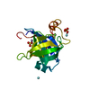
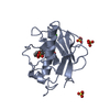
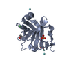
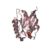
- PDB-3d3d: Bacteriophage lambda lysozyme complexed with a chitohexasaccharide -
+ Open data

- Basic information
| Entry | Database: PDB / ID: 3d3d | |||||||||
|---|---|---|---|---|---|---|---|---|---|---|
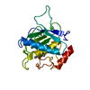
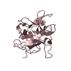
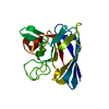
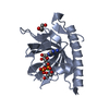
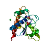
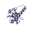
| Title | Bacteriophage lambda lysozyme complexed with a chitohexasaccharide | |||||||||
 Components Components | Lysozyme | |||||||||
 Keywords Keywords | HYDROLASE / GLYCOSIDASE / TRANSGLYCOSYLASE / LYSOZYME / PROTEIN-CHITOHEXASSACHARIDE COMPLEX / Antimicrobial / Bacteriolytic enzyme | |||||||||
| Function / homology |  Function and homology information Function and homology information: / lytic transglycosylase activity / viral release from host cell by cytolysis / peptidoglycan catabolic process / cell wall macromolecule catabolic process / lysozyme activity / host cell cytoplasm / defense response to bacteriumSimilarity search - Function | |||||||||
| Biological species |  Enterobacteria phage lambda (virus) Enterobacteria phage lambda (virus) | |||||||||
| Method | X-RAY DIFFRACTION / MOLECULAR REPLACEMENT / molecular replacement / Resolution: 2.6 Å | |||||||||
 Authors Authors | Leung, A.K.W. / Berghuis, A.M. | |||||||||
 Citation Citation | Journal: Biochemistry / Year: 2001 Title: Crystal structure of the lytic transglycosylase from bacteriophage lambda in complex with hexa-N-acetylchitohexaose Authors: Leung, A.K.W. / Duewel, H.S. / Honek, J.F. / Berghuis, A.M. #1: Journal: Biochim.Biophys.Acta / Year: 1995Title: Investigation of the interactions of saccharides with the lysozyme from bacteriophage lambda Authors: Duewel, H.S. / Daub, E. / Honek, J.F. | |||||||||
| History |
|
- Structure visualization
| Structure viewer | Molecule: MolmilJmol/JSmol |
|---|
- Downloads & links
-Download
| PDBx/mmCIF format | 3d3d.cif.gz | 78.5 KB | Display | PDBx/mmCIF format |
|---|---|---|---|---|
| PDB format | pdb3d3d.ent.gz | 59.4 KB | Display | PDB format |
| PDBx/mmJSON format | 3d3d.json.gz | Tree view | PDBx/mmJSON format | |
| Others |  Other downloads Other downloads |
-Validation report
| Arichive directory | https://data.pdbj.org/pub/pdb/validation_reports/d3/3d3dftp://data.pdbj.org/pub/pdb/validation_reports/d3/3d3d | HTTPS FTP |
|---|
-Related structure data
| Related structure data |  1d9uC 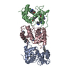 1am7S C: citing same article ( S: Starting model for refinement |
|---|---|
| Similar structure data |
-Links
 PDBj
PDBj


- Assembly
| Deposited unit | 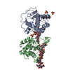
| ||||||||
|---|---|---|---|---|---|---|---|---|---|
| 1 | 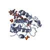
| ||||||||
| 2 | 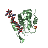
| ||||||||
| Unit cell |
|
-Components
| #1: Protein | / Lysis protein / Muramidase / Endolysin Mass: 17395.701 Da / Num. of mol.: 2 Source method: isolated from a genetically manipulated source Source: (gene. exp.) Enterobacteria phage lambda (virus) / Strain: Lambda / Gene: R / Plasmid: pBR322 / Production host:  Escherichia coli (E. coli) / Strain (production host): TG-1 / References: UniProt: P03706, lysozyme Escherichia coli (E. coli) / Strain (production host): TG-1 / References: UniProt: P03706, lysozyme#2: Polysaccharide | / Mass: 1237.172 Da / Num. of mol.: 2Source method: isolated from a genetically manipulated source #3: Chemical | ChemComp-SO4 / Sulfate  Mass: 96.063 Da / Num. of mol.: 4 / Source method: obtained synthetically / Formula: SO4 Mass: 96.063 Da / Num. of mol.: 4 / Source method: obtained synthetically / Formula: SO4#4: Water | ChemComp-HOH / | Water Mass: 18.015 Da / Num. of mol.: 39 / Source method: isolated from a natural source / Formula: H2O Mass: 18.015 Da / Num. of mol.: 39 / Source method: isolated from a natural source / Formula: H2O |
|---|
-Experimental details
-Experiment
| Experiment | Method: X-RAY DIFFRACTION / Number of used crystals: 1 |
|---|
- Sample preparation
| Crystal | Density Matthews: 2.81 Å3/Da / Density % sol: 56.16 % |
|---|---|
| Crystal grow | Temperature: 298 K / Method: microdialysis / pH: 4.6 Details: 0.1 M NaOAc pH 4.6, 0.1 M ammonium sulfate, 20% w/v PEG 2000 MME, microdialysis, temperature 298K |
-Data collection
| Diffraction | Mean temperature: 100 K |
|---|---|
| Diffraction source | Source: ROTATING ANODE / Type: RIGAKU RU200 / Wavelength: 1.502 Å |
| Detector | Type: RIGAKU RAXIS IIC / Detector: IMAGE PLATE / Date: Aug 30, 1996 / Details: Supper double focusing mirrors |
| Radiation | Protocol: SINGLE WAVELENGTH / Monochromatic (M) / Laue (L): M / Scattering type: x-ray |
| Radiation wavelength | Wavelength: 1.502 Å / Relative weight: 1 |
| Reflection | Resolution: 2.6→33.942 Å / Num. all: 15397 / Num. obs: 11801 / % possible obs: 92 % / Redundancy: 7.7 % / Biso Wilson estimate: 19.3 Å2 / Rmerge(I) obs: 0.118 / Net I/σ(I): 7.9 |
| Reflection shell | Resolution: 2.6→2.76 Å / Rmerge(I) obs: 0.507 / Num. unique all: 947 / % possible all: 51.9 |
-Phasing
| Phasing | Method: molecular replacement |
|---|
- Processing
| Software |
| ||||||||||||||||||||||||||||||||||||
|---|---|---|---|---|---|---|---|---|---|---|---|---|---|---|---|---|---|---|---|---|---|---|---|---|---|---|---|---|---|---|---|---|---|---|---|---|---|
| Refinement | Method to determine structure: MOLECULAR REPLACEMENT Starting model: PDB ENTRY 1AM7 Resolution: 2.6→33.94 Å / Rfactor Rfree error: 0.008 / FOM work R set: 0.814 / Data cutoff high absF: 1696147.25 / Data cutoff low absF: 0 / Isotropic thermal model: RESTRAINED / Cross valid method: THROUGHOUT / σ(F): 0 / Details: BULK SOLVENT MODEL USED
| ||||||||||||||||||||||||||||||||||||
| Solvent computation | Solvent model: FLAT MODEL / Bsol: 7.732 Å2 / ksol: 0.35 e/Å3 | ||||||||||||||||||||||||||||||||||||
| Displacement parameters | Biso mean: 29.4 Å2
| ||||||||||||||||||||||||||||||||||||
| Refine analyze |
| ||||||||||||||||||||||||||||||||||||
| Refinement step | Cycle: LAST / Resolution: 2.6→33.94 Å
| ||||||||||||||||||||||||||||||||||||
| Refine LS restraints |
| ||||||||||||||||||||||||||||||||||||
| LS refinement shell | Resolution: 2.6→2.76 Å / Rfactor Rfree error: 0.032 / Total num. of bins used: 6
| ||||||||||||||||||||||||||||||||||||
| Xplor file |
|
