 Movie
Movie Controller

[English] 日本語
 Yorodumi
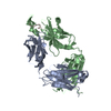
- PDB-3d0v: Crystal structure of the HIV-1 Cross Neutralizing Monoclonal Anti... -
+ Open data

- Basic information
| Entry | Database: PDB / ID: 3d0v | ||||||
|---|---|---|---|---|---|---|---|
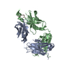
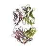
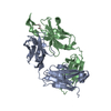
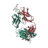
| Title | Crystal structure of the HIV-1 Cross Neutralizing Monoclonal Antibody 2F5 in complex with gp41 Peptide LLELDKWASLW | ||||||
 Components Components |
| ||||||
 Keywords Keywords |  IMMUNE SYSTEM / 2F5 / HIV / gp41 / antibody / neutralizing IMMUNE SYSTEM / 2F5 / HIV / gp41 / antibody / neutralizing | ||||||
| Function / homology |  Function and homology information Function and homology informationvirus-mediated perturbation of host defense response => GO:0019049 / : / Synthesis and processing of ENV and VPU / evasion of host immune response / Alpha-defensins / Dectin-2 family / immunoglobulin complex / Binding and entry of HIV virion / positive regulation of plasma membrane raft polarization / positive regulation of receptor clustering ...virus-mediated perturbation of host defense response => GO:0019049 / : / Synthesis and processing of ENV and VPU / evasion of host immune response / Alpha-defensins / Dectin-2 family / immunoglobulin complex / Binding and entry of HIV virion / positive regulation of plasma membrane raft polarization / positive regulation of receptor clustering / positive regulation of establishment of T cell polarity / virus-mediated perturbation of host defense response / host cell endosome membrane / actin filament organization / Assembly Of The HIV Virion / Budding and maturation of HIV virion / clathrin-dependent endocytosis of virus by host cell / membrane => GO:0016020 / viral protein processing / symbiont entry into host cell / fusion of virus membrane with host plasma membrane / fusion of virus membrane with host endosome membrane / viral envelope / virion attachment to host cell / host cell plasma membrane / virion membrane / structural molecule activity / membrane / plasma membraneSimilarity search - Function | ||||||
| Biological species |  Homo sapiens (human) Homo sapiens (human) | ||||||
| Method | X-RAY DIFFRACTION / MOLECULAR REPLACEMENT / Resolution: 2.05 Å | ||||||
 Authors Authors | Bryson, S. / Julien, J.P. / Pai, E.F. | ||||||
 Citation Citation | Journal: J.Mol.Biol. / Year: 2008 Title: Structural details of HIV-1 recognition by the broadly neutralizing monoclonal antibody 2F5: epitope conformation, antigen-recognition loop mobility, and anion-binding site. Authors: Julien, J.P. / Bryson, S. / Nieva, J.L. / Pai, E.F. | ||||||
| History |
|
- Structure visualization
| Structure viewer | Molecule: MolmilJmol/JSmol |
|---|
- Downloads & links
-Download
| PDBx/mmCIF format | 3d0v.cif.gz | 102.9 KB | Display | PDBx/mmCIF format |
|---|---|---|---|---|
| PDB format | pdb3d0v.ent.gz | 77.6 KB | Display | PDB format |
| PDBx/mmJSON format | 3d0v.json.gz | Tree view | PDBx/mmJSON format | |
| Others |  Other downloads Other downloads |
-Validation report
| Arichive directory | https://data.pdbj.org/pub/pdb/validation_reports/d0/3d0vftp://data.pdbj.org/pub/pdb/validation_reports/d0/3d0v | HTTPS FTP |
|---|
-Related structure data
| Related structure data | 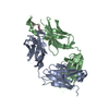 2p8lC 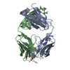 2p8mC 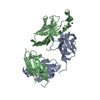 2p8pC 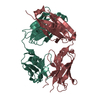 2pr4C 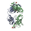 3d0lC 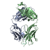 3droC 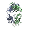 3drqC 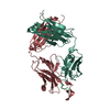 2f5bS C: citing same article ( S: Starting model for refinement |
|---|---|
| Similar structure data |


-Links
 PDBj
PDBj


- Assembly
| Deposited unit | 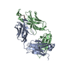
| ||||||||
|---|---|---|---|---|---|---|---|---|---|
| 1 |
| ||||||||
| Unit cell |
|
-Components
| #1: Antibody | Mass: 23363.844 Da / Num. of mol.: 1 / Source method: isolated from a natural source / Source: (natural) Homo sapiens (human) |
|---|---|
| #2: Antibody | Mass: 25245.703 Da / Num. of mol.: 1 / Source method: isolated from a natural source / Source: (natural) Homo sapiens (human) / References: UniProt: Q8NF17*PLUS |
| #3: Protein/peptide | Mass: 1374.603 Da / Num. of mol.: 1 / Source method: obtained synthetically Details: The peptide was chemically synthesized. The sequence of the peptide is naturally found in the human immunodeficiency virus References: UniProt: Q7SVL4, UniProt: P04578*PLUS |
| #4: Water | ChemComp-HOH / Water Mass: 18.015 Da / Num. of mol.: 222 / Source method: isolated from a natural source / Formula: H2O Mass: 18.015 Da / Num. of mol.: 222 / Source method: isolated from a natural source / Formula: H2O |
-Experimental details
-Experiment
| Experiment | Method: X-RAY DIFFRACTION / Number of used crystals: 1 |
|---|
- Sample preparation
| Crystal | Density Matthews: 3.34 Å3/Da / Density % sol: 63.19 % |
|---|---|
| Crystal grow | Temperature: 293 K / Method: vapor diffusion, hanging drop / pH: 5.6 Details: 0.1M sodium acetate, pH 5.6, 20% 2-propanol, 16-20% PEG 4000, VAPOR DIFFUSION, HANGING DROP, temperature 293K |
-Data collection
| Diffraction | Mean temperature: 100 K |
|---|---|
| Diffraction source | Source: ROTATING ANODE / Type: RIGAKU / Wavelength: 1.54 Å |
| Detector | Type: MAR scanner 345 mm plate / Detector: IMAGE PLATE / Date: Jul 7, 1999 |
| Radiation | Protocol: SINGLE WAVELENGTH / Monochromatic (M) / Laue (L): M / Scattering type: x-ray |
| Radiation wavelength | Wavelength: 1.54 Å / Relative weight: 1 |
| Reflection | Resolution: 2.03→20 Å / Num. all: 44152 / Num. obs: 42170 / % possible obs: 95.5 % / Biso Wilson estimate: 25.7 Å2 / Rsym value: 0.058 |
| Reflection shell | Resolution: 2.05→2.18 Å / Num. unique all: 6083 / Rsym value: 0.3 |
- Processing
| Software |
| ||||||||||||||||||||||||||||||||||||||||||||||||||||||||||||
|---|---|---|---|---|---|---|---|---|---|---|---|---|---|---|---|---|---|---|---|---|---|---|---|---|---|---|---|---|---|---|---|---|---|---|---|---|---|---|---|---|---|---|---|---|---|---|---|---|---|---|---|---|---|---|---|---|---|---|---|---|---|
| Refinement | Method to determine structure: MOLECULAR REPLACEMENT Starting model: 2F5B Resolution: 2.05→19.76 Å / Rfactor Rfree error: 0.005 / Data cutoff high absF: 965007.6 / Data cutoff low absF: 0 / Isotropic thermal model: OVERALL / Cross valid method: THROUGHOUT / σ(F): 0
| ||||||||||||||||||||||||||||||||||||||||||||||||||||||||||||
| Solvent computation | Solvent model: FLAT MODEL / Bsol: 56.7935 Å2 / ksol: 0.406975 e/Å3 | ||||||||||||||||||||||||||||||||||||||||||||||||||||||||||||
| Displacement parameters | Biso mean: 37.1 Å2
| ||||||||||||||||||||||||||||||||||||||||||||||||||||||||||||
| Refine analyze |
| ||||||||||||||||||||||||||||||||||||||||||||||||||||||||||||
| Refinement step | Cycle: LAST / Resolution: 2.05→19.76 Å
| ||||||||||||||||||||||||||||||||||||||||||||||||||||||||||||
| Refine LS restraints |
| ||||||||||||||||||||||||||||||||||||||||||||||||||||||||||||
| LS refinement shell | Resolution: 2.05→2.18 Å / Rfactor Rfree error: 0.015 / Total num. of bins used: 6
| ||||||||||||||||||||||||||||||||||||||||||||||||||||||||||||
| Xplor file |
|
