 Movie
Movie Controller

[English] 日本語
 Yorodumi
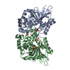
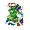
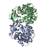
- PDB-3d0c: Crystal structure of dihydrodipicolinate synthase from Oceanobaci... -
+ Open data

- Basic information
| Entry | Database: PDB / ID: 3d0c | ||||||
|---|---|---|---|---|---|---|---|
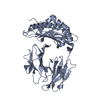
| Title | Crystal structure of dihydrodipicolinate synthase from Oceanobacillus iheyensis at 1.9 A resolution | ||||||
 Components Components | Dihydrodipicolinate synthase | ||||||
 Keywords Keywords | LYASE / Lysine biosynthesis / Pyruvate / TIM barrel / NYSGXRC / 9375o / PSI-2 / Structural Genomics / Protein Structure Initiative / New York SGX Research Center for Structural Genomics | ||||||
| Function / homology |  Function and homology information4-hydroxy-tetrahydrodipicolinate synthase / 4-hydroxy-tetrahydrodipicolinate synthase activity Function and homology information4-hydroxy-tetrahydrodipicolinate synthase / 4-hydroxy-tetrahydrodipicolinate synthase activitySimilarity search - Function | ||||||
| Biological species |  Oceanobacillus iheyensis HTE831 (bacteria) Oceanobacillus iheyensis HTE831 (bacteria) | ||||||
| Method | X-RAY DIFFRACTION / SYNCHROTRON / SAD / Resolution: 1.9 Å | ||||||
 Authors Authors | Satyanarayana, L. / Eswaramoorthy, S. / Sauder, J.M. / Burley, S.K. / Swaminathan, S. / New York SGX Research Center for Structural Genomics (NYSGXRC) | ||||||
 Citation Citation | Journal: To be Published Title: Crystal structure of dihydrodipicolinate synthase from Oceanobacillus iheyensis at 1.9 A resolution. Authors: Satyanarayana, L. / Eswaramoorthy, S. / Sauder, J.M. / Burley, S.K. / Swaminathan, S. | ||||||
| History |
|
- Structure visualization
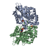
| Structure viewer | Molecule: MolmilJmol/JSmol |
|---|
- Downloads & links
-Download
| PDBx/mmCIF format | 3d0c.cif.gz | 132.9 KB | Display | PDBx/mmCIF format |
|---|---|---|---|---|
| PDB format | pdb3d0c.ent.gz | 108.2 KB | Display | PDB format |
| PDBx/mmJSON format | 3d0c.json.gz | Tree view | PDBx/mmJSON format | |
| Others |  Other downloads Other downloads |
-Validation report
| Arichive directory | https://data.pdbj.org/pub/pdb/validation_reports/d0/3d0cftp://data.pdbj.org/pub/pdb/validation_reports/d0/3d0c | HTTPS FTP |
|---|
-Related structure data
| Similar structure data | |
|---|---|
| Other databases |

-Links
 PDBj
PDBj- Assembly
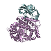
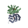
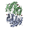
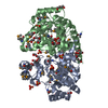
| Deposited unit | 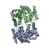
| ||||||||
|---|---|---|---|---|---|---|---|---|---|
| 1 |
| ||||||||
| Unit cell |
| ||||||||
| Details | AUTHORS STATE THAT THE DIMERIC ASSEMBLY OF THE BIOLOGICAL UNIT THAT IS SHOWN IN REMARK 350 IS PUTATIVE. |
-Components
| #1: Protein | Mass: 35149.949 Da / Num. of mol.: 2 Source method: isolated from a genetically manipulated source Source: (gene. exp.) Oceanobacillus iheyensis HTE831 (bacteria)Species: Oceanobacillus iheyensis / Strain: HTE831 / DSM 14371 / JCM 11309 / KCTC 3954 / / Gene: OB2845, DHDPS / Plasmid: BC-pSGX3(BC) / Production host: Escherichia coli (E. coli) / Strain (production host): BL21(DE3)Codon+RIL / References: UniProt: Q8EMJ7, dihydrodipicolinate synthase#2: Water | ChemComp-HOH / | Water Mass: 18.015 Da / Num. of mol.: 442 / Source method: isolated from a natural source / Formula: H2O Mass: 18.015 Da / Num. of mol.: 442 / Source method: isolated from a natural source / Formula: H2O |
|---|
-Experimental details
-Experiment
| Experiment | Method: X-RAY DIFFRACTION / Number of used crystals: 1 |
|---|
- Sample preparation
| Crystal | Density Matthews: 2.28 Å3/Da / Density % sol: 45.98 % |
|---|---|
| Crystal grow | Temperature: 293 K / Method: vapor diffusion, sitting drop / pH: 8.5 Details: 0.2M Magnesium chloride, 0.1M Lysine, 3% DMSO, 0.1M Tris-HCl pH 8.5, 25% PEG 3350, VAPOR DIFFUSION, SITTING DROP, temperature 293K |
-Data collection
| Diffraction | Mean temperature: 100 K |
|---|---|
| Diffraction source | Source: SYNCHROTRON / Site: NSLS  / Beamline: X29A / Wavelength: 0.9792 Å / Beamline: X29A / Wavelength: 0.9792 Å |
| Detector | Type: ADSC QUANTUM 315 / Detector: CCD / Date: Apr 21, 2008 / Details: Mirrors |
| Radiation | Monochromator: Si 111 CHANNEL / Protocol: SINGLE WAVELENGTH / Monochromatic (M) / Laue (L): M / Scattering type: x-ray |
| Radiation wavelength | Wavelength: 0.9792 Å / Relative weight: 1 |
| Reflection | Resolution: 1.9→50 Å / Num. all: 50112 / Num. obs: 50112 / % possible obs: 96.1 % / Observed criterion σ(F): 0 / Observed criterion σ(I): 0 / Redundancy: 11.3 % / Biso Wilson estimate: 17.5 Å2 / Rmerge(I) obs: 0.074 / Net I/σ(I): 10.4 |
| Reflection shell | Resolution: 1.9→1.97 Å / Redundancy: 8.2 % / Rmerge(I) obs: 0.35 / Num. unique all: 4270 / % possible all: 85 |
- Processing
| Software |
| |||||||||||||||||||||||||
|---|---|---|---|---|---|---|---|---|---|---|---|---|---|---|---|---|---|---|---|---|---|---|---|---|---|---|
| Refinement | Method to determine structure: SAD / Resolution: 1.9→50 Å / Isotropic thermal model: Isotropic / Cross valid method: THROUGHOUT / Stereochemistry target values: Engh & Huber Details: Residues listed as missing in Remark 465 are due to lack of electron density. Residues with missing atoms listed in Remark 470 are due to lack of electron density for side chains and modeled as alanines.
| |||||||||||||||||||||||||
| Displacement parameters | Biso mean: 27.4 Å2
| |||||||||||||||||||||||||
| Refine analyze |
| |||||||||||||||||||||||||
| Refinement step | Cycle: LAST / Resolution: 1.9→50 Å
| |||||||||||||||||||||||||
| Refine LS restraints |
| |||||||||||||||||||||||||
| LS refinement shell | Resolution: 1.9→2.02 Å / Rfactor Rfree error: 0.016
|
