 Movie
Movie Controller


+ Open data

- Basic information
| Entry | Database: PDB / ID: 3cun | ||||||
|---|---|---|---|---|---|---|---|
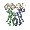
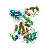
| Title | Aminoacyl-tRNA synthetase ribozyme | ||||||
 Components Components |
| ||||||
 Keywords Keywords | RNA/RNA binding / ARS ribozyme catalytic RNA / RNA-RNA binding COMPLEX | ||||||
| Function / homology |  Function and homology information Function and homology informationU1 snRNP binding / U1 snRNP /  U1 snRNA binding / U4/U6 x U5 tri-snRNP complex / mRNA Splicing - Major Pathway / spliceosomal complex / mRNA splicing, via spliceosome / DNA binding / RNA binding / nucleoplasm ...U1 snRNP binding / U1 snRNP / U1 snRNA binding / U4/U6 x U5 tri-snRNP complex / mRNA Splicing - Major Pathway / spliceosomal complex / mRNA splicing, via spliceosome / DNA binding / RNA binding / nucleoplasm / identical protein binding / nucleus U1 snRNA binding / U4/U6 x U5 tri-snRNP complex / mRNA Splicing - Major Pathway / spliceosomal complex / mRNA splicing, via spliceosome / DNA binding / RNA binding / nucleoplasm ...U1 snRNP binding / U1 snRNP / U1 snRNA binding / U4/U6 x U5 tri-snRNP complex / mRNA Splicing - Major Pathway / spliceosomal complex / mRNA splicing, via spliceosome / DNA binding / RNA binding / nucleoplasm / identical protein binding / nucleusSimilarity search - Function | ||||||
| Biological species |  Homo sapiens (human) Homo sapiens (human) | ||||||
| Method | X-RAY DIFFRACTION / SYNCHROTRON / FOURIER SYNTHESIS / Resolution: 3 Å | ||||||
 Authors Authors | Xiao, H. / Murakami, H. / Suga, H. / Ferre-D'Amare, A.R. | ||||||
 Citation Citation | Journal: Nature / Year: 2008 Title: Structural basis of specific tRNA aminoacylation by a small in vitro selected ribozyme. Authors: Xiao, H. / Murakami, H. / Suga, H. / Ferre-D'Amare, A.R. | ||||||
| History |
|
- Structure visualization
| Structure viewer | Molecule: MolmilJmol/JSmol |
|---|
- Downloads & links
-Download
| PDBx/mmCIF format | 3cun.cif.gz | 151.5 KB | Display | PDBx/mmCIF format |
|---|---|---|---|---|
| PDB format | pdb3cun.ent.gz | 112.8 KB | Display | PDB format |
| PDBx/mmJSON format | 3cun.json.gz | Tree view | PDBx/mmJSON format | |
| Others |  Other downloads Other downloads |
-Validation report
| Arichive directory | https://data.pdbj.org/pub/pdb/validation_reports/cu/3cunftp://data.pdbj.org/pub/pdb/validation_reports/cu/3cun | HTTPS FTP |
|---|
-Related structure data
| Related structure data | 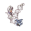 3culSC S: Starting model for refinement C: citing same article ( |
|---|---|
| Similar structure data |


-Links
 PDBj
PDBj


- Assembly
| Deposited unit | 
| ||||||||
|---|---|---|---|---|---|---|---|---|---|
| 1 | 
| ||||||||
| 2 | 
| ||||||||
| Unit cell |
|
-Components
| #1: RNA chain | Mass: 29820.535 Da / Num. of mol.: 2 Source method: isolated from a genetically manipulated source Details: Flexizyme / Production host: CELL-FREE SYNTHESIS (others) #2: Protein | Mass: 11574.792 Da / Num. of mol.: 2 / Fragment: UNP residues 1-98 / Mutation: Y31H, Q36R Source method: isolated from a genetically manipulated source Source: (gene. exp.) Homo sapiens (human) / Gene: SNRPA / Production host:  Escherichia coli (E. coli) / References: UniProt: P09012 Escherichia coli (E. coli) / References: UniProt: P09012#3: Chemical | ChemComp-MG /   Mass: 24.305 Da / Num. of mol.: 9 / Source method: obtained synthetically / Formula: Mg Mass: 24.305 Da / Num. of mol.: 9 / Source method: obtained synthetically / Formula: Mg#4: Chemical | ChemComp-K / |   Mass: 39.098 Da / Num. of mol.: 1 / Source method: obtained synthetically / Formula: K Mass: 39.098 Da / Num. of mol.: 1 / Source method: obtained synthetically / Formula: K#5: Chemical | ChemComp-CO / |   Mass: 58.933 Da / Num. of mol.: 1 / Source method: obtained synthetically / Formula: Co Mass: 58.933 Da / Num. of mol.: 1 / Source method: obtained synthetically / Formula: Co |
|---|
-Experimental details
-Experiment
| Experiment | Method: X-RAY DIFFRACTION / Number of used crystals: 1 |
|---|
- Sample preparation
| Crystal | Density Matthews: 2.51 Å3/Da / Density % sol: 51.07 % | ||||||||||||||||||||
|---|---|---|---|---|---|---|---|---|---|---|---|---|---|---|---|---|---|---|---|---|---|
| Crystal grow | Temperature: 295 K / Method: vapor diffusion / pH: 7 Details: 100 mM magenesium formate pH 7.0, 15% PEG 3000, 1 M lithium chloride, VAPOR DIFFUSION, temperature 295K | ||||||||||||||||||||
| Components of the solutions |
|
-Data collection
| Diffraction | Mean temperature: 100 K |
|---|---|
| Diffraction source | Source: SYNCHROTRON / Site: ALS  / Beamline: 5.0.2 / Wavelength: 0.9572 Å / Beamline: 5.0.2 / Wavelength: 0.9572 Å |
| Detector | Type: ADSC QUANTUM 315 / Detector: CCD / Date: Oct 6, 2005 / Details: Si (111) |
| Radiation | Monochromator: Si (111) / Protocol: SINGLE WAVELENGTH / Monochromatic (M) / Laue (L): M / Scattering type: x-ray |
| Radiation wavelength | Wavelength: 0.9572 Å / Relative weight: 1 |
| Reflection | Resolution: 3→30 Å / Num. obs: 13610 / % possible obs: 80.8 % / Observed criterion σ(I): 1.9 / Redundancy: 6.4 % / Rmerge(I) obs: 0.87 / Rsym value: 0.87 / Net I/σ(I): 21.1 |
| Reflection shell | Resolution: 3→3.1 Å / Redundancy: 2.9 % / Rmerge(I) obs: 0.501 / Mean I/σ(I) obs: 1.9 / Num. unique all: 1355 / Rsym value: 0.501 / % possible all: 51.6 |
- Processing
| Software |
| ||||||||||||||||||||||||||||||||||||
|---|---|---|---|---|---|---|---|---|---|---|---|---|---|---|---|---|---|---|---|---|---|---|---|---|---|---|---|---|---|---|---|---|---|---|---|---|---|
| Refinement | Method to determine structure: FOURIER SYNTHESIS Starting model: PDB entry 3CUL Resolution: 3→28.75 Å / Rfactor Rfree error: 0.008 / Data cutoff high absF: 185743.74 / Data cutoff low absF: 0 / Isotropic thermal model: RESTRAINED / Cross valid method: THROUGHOUT / σ(F): 0 / Stereochemistry target values: Engh & Huber
| ||||||||||||||||||||||||||||||||||||
| Solvent computation | Solvent model: FLAT MODEL / Bsol: 25 Å2 / ksol: 0.3 e/Å3 | ||||||||||||||||||||||||||||||||||||
| Displacement parameters | Biso mean: 100.7 Å2
| ||||||||||||||||||||||||||||||||||||
| Refine analyze |
| ||||||||||||||||||||||||||||||||||||
| Refinement step | Cycle: LAST / Resolution: 3→28.75 Å
| ||||||||||||||||||||||||||||||||||||
| Refine LS restraints |
| ||||||||||||||||||||||||||||||||||||
| LS refinement shell | Resolution: 3→3.19 Å / Rfactor Rfree error: 0.038 / Total num. of bins used: 6
| ||||||||||||||||||||||||||||||||||||
| Xplor file |
|
