 Movie
Movie Controller


+ Open data

- Basic information
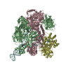
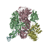
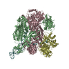
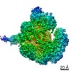
| Entry | Database: PDB / ID: 3cr8 | ||||||
|---|---|---|---|---|---|---|---|
| Title | Hexameric APS kinase from Thiobacillus denitrificans | ||||||
 Components Components | Sulfate adenylyltransferase, adenylylsulfate kinase | ||||||
 Keywords Keywords | TRANSFERASE / APS kinase / Adenylylsulfate kinase / sulfate metabolism / Nucleotide 2 kinase / Nucleotidyltransferase | ||||||
| Function / homology |  Function and homology informationadenylyl-sulfate kinase / adenylylsulfate kinase activity / sulfate adenylyltransferase (ATP) activity / sulfate assimilation / hydrogen sulfide biosynthetic process / ATP binding Function and homology informationadenylyl-sulfate kinase / adenylylsulfate kinase activity / sulfate adenylyltransferase (ATP) activity / sulfate assimilation / hydrogen sulfide biosynthetic process / ATP bindingSimilarity search - Function | ||||||
| Biological species |  Thiobacillus denitrificans (bacteria) Thiobacillus denitrificans (bacteria) | ||||||
| Method | X-RAY DIFFRACTION / SYNCHROTRON / MOLECULAR REPLACEMENT / molecular replacement / Resolution: 2.95 Å | ||||||
 Authors Authors | Gay, S.C. / Segel, I.H. / Fisher, A.J. | ||||||
 Citation Citation | Journal: Acta Crystallogr.,Sect.D / Year: 2009 Title: Structure of the two-domain hexameric APS kinase from Thiobacillus denitrificans: structural basis for the absence of ATP sulfurylase activity. Authors: Gay, S.C. / Segel, I.H. / Fisher, A.J. | ||||||
| History |
|
- Structure visualization
| Structure viewer | Molecule: MolmilJmol/JSmol |
|---|
- Downloads & links
-Download
| PDBx/mmCIF format | 3cr8.cif.gz | 291 KB | Display | PDBx/mmCIF format |
|---|---|---|---|---|
| PDB format | pdb3cr8.ent.gz | 234.4 KB | Display | PDB format |
| PDBx/mmJSON format | 3cr8.json.gz | Tree view | PDBx/mmJSON format | |
| Others |  Other downloads Other downloads |
-Validation report
| Arichive directory | https://data.pdbj.org/pub/pdb/validation_reports/cr/3cr8ftp://data.pdbj.org/pub/pdb/validation_reports/cr/3cr8 | HTTPS FTP |
|---|
-Related structure data
| Related structure data |  1i2dS S: Starting model for refinement |
|---|---|
| Similar structure data |


-Links
 PDBj
PDBj
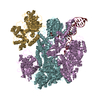
- Assembly
| Deposited unit | 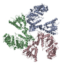
| |||||||||||||||||||||||||||||||||||||||||||||||||||||||||||||||||||||||||||||||||||||||||||||||||||||||||||||||||||||
|---|---|---|---|---|---|---|---|---|---|---|---|---|---|---|---|---|---|---|---|---|---|---|---|---|---|---|---|---|---|---|---|---|---|---|---|---|---|---|---|---|---|---|---|---|---|---|---|---|---|---|---|---|---|---|---|---|---|---|---|---|---|---|---|---|---|---|---|---|---|---|---|---|---|---|---|---|---|---|---|---|---|---|---|---|---|---|---|---|---|---|---|---|---|---|---|---|---|---|---|---|---|---|---|---|---|---|---|---|---|---|---|---|---|---|---|---|---|---|
| 1 | 
| |||||||||||||||||||||||||||||||||||||||||||||||||||||||||||||||||||||||||||||||||||||||||||||||||||||||||||||||||||||
| Unit cell |
| |||||||||||||||||||||||||||||||||||||||||||||||||||||||||||||||||||||||||||||||||||||||||||||||||||||||||||||||||||||
| Components on special symmetry positions |
| |||||||||||||||||||||||||||||||||||||||||||||||||||||||||||||||||||||||||||||||||||||||||||||||||||||||||||||||||||||
| Noncrystallographic symmetry (NCS) | NCS domain:
NCS domain segments: Ens-ID: 1
|
-Components
| #1: Protein | Mass: 61638.320 Da / Num. of mol.: 3 Source method: isolated from a genetically manipulated source Source: (gene. exp.) Thiobacillus denitrificans (bacteria) / Strain: ATCC 25259 / Plasmid: pET22b / Production host: Escherichia coli (E. coli) / Strain (production host): BL21(DE3)References: UniProt: Q3SM86, sulfate adenylyltransferase, adenylyl-sulfate kinase#2: Water | ChemComp-HOH / | Water Mass: 18.015 Da / Num. of mol.: 86 / Source method: isolated from a natural source / Formula: H2O Mass: 18.015 Da / Num. of mol.: 86 / Source method: isolated from a natural source / Formula: H2O |
|---|
-Experimental details
-Experiment
| Experiment | Method: X-RAY DIFFRACTION / Number of used crystals: 1 |
|---|
- Sample preparation
| Crystal | Density Matthews: 2.64 Å3/Da / Density % sol: 53.43 % |
|---|---|
| Crystal grow | Temperature: 293 K / Method: vapor diffusion, hanging drop / pH: 7 Details: K/Na Tartrate, NaCl, Bis Tris Propane, pH 7, VAPOR DIFFUSION, HANGING DROP, temperature 293K |
-Data collection
| Diffraction | Mean temperature: 100 K |
|---|---|
| Diffraction source | Source: SYNCHROTRON / Site: SSRL  / Beamline: BL9-2 / Wavelength: 0.979 Å / Beamline: BL9-2 / Wavelength: 0.979 Å |
| Detector | Type: ADSC QUANTUM 315 / Detector: CCD / Date: Feb 28, 2007 / Details: double crystal monochromator |
| Radiation | Monochromator: Double Si(111) crystal monochromator / Protocol: SINGLE WAVELENGTH / Monochromatic (M) / Laue (L): M / Scattering type: x-ray |
| Radiation wavelength | Wavelength: 0.979 Å / Relative weight: 1 |
| Reflection | Resolution: 2.95→50 Å / Num. all: 3317 / Num. obs: 38467 / % possible obs: 92.4 % / Observed criterion σ(F): -3 / Observed criterion σ(I): -3 / Redundancy: 3 % / Rmerge(I) obs: 0.107 / Χ2: 1.084 / Net I/σ(I): 7.3 |
| Reflection shell | Resolution: 2.95→3.06 Å / Redundancy: 3.3 % / Rmerge(I) obs: 0.357 / Num. unique all: 3925 / Χ2: 0.818 / % possible all: 95.8 |
-Phasing
| Phasing | Method: molecular replacement | ||||||
|---|---|---|---|---|---|---|---|
| Phasing MR |
|
- Processing
| Software |
| ||||||||||||||||||||||||||||||||||||||||||||||||||||||||||||||||||||||||||||||||||||||||||||||||||
|---|---|---|---|---|---|---|---|---|---|---|---|---|---|---|---|---|---|---|---|---|---|---|---|---|---|---|---|---|---|---|---|---|---|---|---|---|---|---|---|---|---|---|---|---|---|---|---|---|---|---|---|---|---|---|---|---|---|---|---|---|---|---|---|---|---|---|---|---|---|---|---|---|---|---|---|---|---|---|---|---|---|---|---|---|---|---|---|---|---|---|---|---|---|---|---|---|---|---|---|
| Refinement | Method to determine structure: MOLECULAR REPLACEMENT Starting model: PDB entry 1I2D Resolution: 2.95→35.028 Å / Isotropic thermal model: isotropic / σ(F): 1.34 / Stereochemistry target values: ML
| ||||||||||||||||||||||||||||||||||||||||||||||||||||||||||||||||||||||||||||||||||||||||||||||||||
| Solvent computation | Shrinkage radii: 0.9 Å / VDW probe radii: 1.11 Å / Solvent model: FLAT BULK SOLVENT MODEL | ||||||||||||||||||||||||||||||||||||||||||||||||||||||||||||||||||||||||||||||||||||||||||||||||||
| Displacement parameters | Biso max: 19.07 Å2 / Biso mean: 60.52 Å2 / Biso min: 139.13 Å2
| ||||||||||||||||||||||||||||||||||||||||||||||||||||||||||||||||||||||||||||||||||||||||||||||||||
| Refinement step | Cycle: LAST / Resolution: 2.95→35.028 Å
| ||||||||||||||||||||||||||||||||||||||||||||||||||||||||||||||||||||||||||||||||||||||||||||||||||
| Refine LS restraints |
| ||||||||||||||||||||||||||||||||||||||||||||||||||||||||||||||||||||||||||||||||||||||||||||||||||
| Refine LS restraints NCS |
| ||||||||||||||||||||||||||||||||||||||||||||||||||||||||||||||||||||||||||||||||||||||||||||||||||
| LS refinement shell | Refine-ID: X-RAY DIFFRACTION / Total num. of bins used: 13
|
