 Movie
Movie Controller

[English] 日本語
 Yorodumi
- PDB-3cqn: Crystal Structure of the Lipocalin domain of Violaxanthin de-epox... -
+ Open data

- Basic information
| Entry | Database: PDB / ID: 3cqn | ||||||
|---|---|---|---|---|---|---|---|
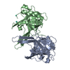
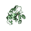
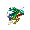
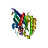
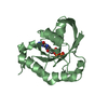
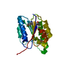
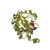
| Title | Crystal Structure of the Lipocalin domain of Violaxanthin de-epoxidase (VDE) at pH7 | ||||||
 Components Components | Violaxanthin de-epoxidase, chloroplast | ||||||
 Keywords Keywords | OXIDOREDUCTASE / Lipocalin / Enzyme / de-epoxidase / Xanthophyll cycle / non photochemical quenching / NPQ / Violaxanthin / Antheraxanthin / Zeaxanthin / pH dependant transition / Chloroplast / Membrane / Thylakoid / Transit peptide | ||||||
| Function / homology |  Function and homology informationviolaxanthin de-epoxidase / xanthophyll cycle / violaxanthin de-epoxidase activity / chlorophyll metabolic process / thylakoid lumen / chloroplast thylakoid / chloroplast thylakoid membrane / chloroplast / fatty acid metabolic process / response to heat ...violaxanthin de-epoxidase / xanthophyll cycle / violaxanthin de-epoxidase activity / chlorophyll metabolic process / thylakoid lumen / chloroplast thylakoid / chloroplast thylakoid membrane / chloroplast / fatty acid metabolic process / response to heat / protein domain specific binding / extracellular region / cytosol Function and homology informationviolaxanthin de-epoxidase / xanthophyll cycle / violaxanthin de-epoxidase activity / chlorophyll metabolic process / thylakoid lumen / chloroplast thylakoid / chloroplast thylakoid membrane / chloroplast / fatty acid metabolic process / response to heat ...violaxanthin de-epoxidase / xanthophyll cycle / violaxanthin de-epoxidase activity / chlorophyll metabolic process / thylakoid lumen / chloroplast thylakoid / chloroplast thylakoid membrane / chloroplast / fatty acid metabolic process / response to heat / protein domain specific binding / extracellular region / cytosolSimilarity search - Function | ||||||
| Biological species |  Arabidopsis thaliana (thale cress) Arabidopsis thaliana (thale cress) | ||||||
| Method | X-RAY DIFFRACTION / SYNCHROTRON / MOLECULAR REPLACEMENT / molecular replacement / Resolution: 2 Å | ||||||
 Authors Authors | Arnoux, P. / Morosinotto, T. / Pignol, D. | ||||||
 Citation Citation | Journal: Plant Cell / Year: 2009 Title: A structural basis for the pH-dependent xanthophyll cycle in Arabidopsis thaliana. Authors: Arnoux, P. / Morosinotto, T. / Saga, G. / Bassi, R. / Pignol, D. | ||||||
| History |
|
- Structure visualization
| Structure viewer | Molecule: MolmilJmol/JSmol |
|---|
- Downloads & links
-Download
| PDBx/mmCIF format | 3cqn.cif.gz | 81 KB | Display | PDBx/mmCIF format |
|---|---|---|---|---|
| PDB format | pdb3cqn.ent.gz | 60.6 KB | Display | PDB format |
| PDBx/mmJSON format | 3cqn.json.gz | Tree view | PDBx/mmJSON format | |
| Others |  Other downloads Other downloads |
-Validation report
| Arichive directory | https://data.pdbj.org/pub/pdb/validation_reports/cq/3cqnftp://data.pdbj.org/pub/pdb/validation_reports/cq/3cqn | HTTPS FTP |
|---|
-Related structure data


-Links
 PDBj
PDBj

- Assembly
| Deposited unit | 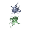
| ||||||||||||||||||
|---|---|---|---|---|---|---|---|---|---|---|---|---|---|---|---|---|---|---|---|
| 1 | 
| ||||||||||||||||||
| 2 | 
| ||||||||||||||||||
| Unit cell |
| ||||||||||||||||||
| Noncrystallographic symmetry (NCS) | NCS domain:
NCS domain segments: Component-ID: 1 / Ens-ID: 1 / Beg auth comp-ID: PRO / Beg label comp-ID: PRO / End auth comp-ID: GLY / End label comp-ID: GLY / Refine code: 4 / Auth seq-ID: 83 - 250 / Label seq-ID: 7 - 174
| ||||||||||||||||||
| Details | biological unit is a monomer at pH7 and a dimer at pH5. There are two biological units in the asymmetric unit at pH7 (chain A and chain B) |
-Components
| #1: Protein | / Protein NON-PHOTOCHEMICAL QUENCHING 1 / AtVxDE Mass: 21343.457 Da / Num. of mol.: 2 / Fragment: Lipocalin Domain (UNP residues 191-366) Source method: isolated from a genetically manipulated source Source: (gene. exp.) Arabidopsis thaliana (thale cress) / Gene: VDE1, AVDE1, NPQ1, VXDE / Plasmid: pQE60 / Production host:  Escherichia coli (E. coli) / Strain (production host): BL21(DE3) / References: UniProt: Q39249, EC: 1.10.99.3 Escherichia coli (E. coli) / Strain (production host): BL21(DE3) / References: UniProt: Q39249, EC: 1.10.99.3#2: Water | ChemComp-HOH / | Water Mass: 18.015 Da / Num. of mol.: 158 / Source method: isolated from a natural source / Formula: H2O Mass: 18.015 Da / Num. of mol.: 158 / Source method: isolated from a natural source / Formula: H2O |
|---|
-Experimental details
-Experiment
| Experiment | Method: X-RAY DIFFRACTION / Number of used crystals: 1 |
|---|
- Sample preparation
| Crystal | Density Matthews: 2.36 Å3/Da / Density % sol: 43.27 % |
|---|---|
| Crystal grow | Temperature: 298 K / Method: vapor diffusion / pH: 7 Details: PEG3350, MgNO3, Benzamidine HCl, pH 7, vapor diffusion, temperature 298K |
-Data collection
| Diffraction | Mean temperature: 100 K |
|---|---|
| Diffraction source | Source: SYNCHROTRON / Site: ESRF  / Beamline: ID23-1 / Wavelength: 0.97625 Å / Beamline: ID23-1 / Wavelength: 0.97625 Å |
| Detector | Type: ADSC QUANTUM 315 / Detector: CCD / Date: Mar 19, 2006 |
| Radiation | Protocol: SINGLE WAVELENGTH / Monochromatic (M) / Laue (L): M / Scattering type: x-ray |
| Radiation wavelength | Wavelength: 0.97625 Å / Relative weight: 1 |
| Reflection | Resolution: 2→30 Å / Num. all: 23574 / Num. obs: 23574 / % possible obs: 96.9 % / Observed criterion σ(F): 1 / Observed criterion σ(I): 1 / Redundancy: 2 % / Biso Wilson estimate: 20.7 Å2 / Rmerge(I) obs: 0.088 / Rsym value: 0.088 / Net I/σ(I): 6.2 |
| Reflection shell | Resolution: 2→2.11 Å / Redundancy: 2 % / Rmerge(I) obs: 0.344 / Mean I/σ(I) obs: 1.6 / Num. unique all: 6787 / Rsym value: 0.344 / % possible all: 97.1 |
-Phasing
| Phasing | Method: molecular replacement |
|---|
- Processing
| Software |
| ||||||||||||||||||||||||||||||||||||||||||||||||||||||||||||||||||||||||||||||||||||||||||
|---|---|---|---|---|---|---|---|---|---|---|---|---|---|---|---|---|---|---|---|---|---|---|---|---|---|---|---|---|---|---|---|---|---|---|---|---|---|---|---|---|---|---|---|---|---|---|---|---|---|---|---|---|---|---|---|---|---|---|---|---|---|---|---|---|---|---|---|---|---|---|---|---|---|---|---|---|---|---|---|---|---|---|---|---|---|---|---|---|---|---|---|
| Refinement | Method to determine structure: MOLECULAR REPLACEMENT Starting model: AtVDE at pH5 Resolution: 2→30 Å / Cor.coef. Fo:Fc: 0.952 / Cor.coef. Fo:Fc free: 0.903 / SU B: 9.915 / SU ML: 0.139 / Cross valid method: THROUGHOUT / σ(F): 0 / ESU R: 0.201 / ESU R Free: 0.197 / Stereochemistry target values: MAXIMUM LIKELIHOOD
| ||||||||||||||||||||||||||||||||||||||||||||||||||||||||||||||||||||||||||||||||||||||||||
| Solvent computation | Ion probe radii: 0.8 Å / Shrinkage radii: 0.8 Å / VDW probe radii: 1.2 Å / Solvent model: MASK | ||||||||||||||||||||||||||||||||||||||||||||||||||||||||||||||||||||||||||||||||||||||||||
| Displacement parameters | Biso mean: 34.02 Å2
| ||||||||||||||||||||||||||||||||||||||||||||||||||||||||||||||||||||||||||||||||||||||||||
| Refinement step | Cycle: LAST / Resolution: 2→30 Å
| ||||||||||||||||||||||||||||||||||||||||||||||||||||||||||||||||||||||||||||||||||||||||||
| Refine LS restraints |
| ||||||||||||||||||||||||||||||||||||||||||||||||||||||||||||||||||||||||||||||||||||||||||
| Refine LS restraints NCS | Dom-ID: 1 / Auth asym-ID: A / Ens-ID: 1 / Number: 1278 / Refine-ID: X-RAY DIFFRACTION
| ||||||||||||||||||||||||||||||||||||||||||||||||||||||||||||||||||||||||||||||||||||||||||
| LS refinement shell | Resolution: 2→2.052 Å / Total num. of bins used: 20
|
