 Movie
Movie Controller

[English] 日本語
 Yorodumi
- PDB-3co3: X-Ray Crystal Structure of a Monofunctional Platinum-DNA Adduct, ... -
+ Open data

- Basic information
| Entry | Database: PDB / ID: 3co3 | ||||||
|---|---|---|---|---|---|---|---|
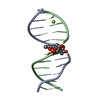
| Title | X-Ray Crystal Structure of a Monofunctional Platinum-DNA Adduct, cis-{Pt(NH3)2(pyridine)}2+ Bound to Deoxyguanosine in a Dodecamer Duplex | ||||||
 Components Components |
| ||||||
 Keywords Keywords |  DNA / PLATINUM-DNA DUPLEX / CISPLATIN / MONOFUNCTIONAL PT COMPOUND DNA / PLATINUM-DNA DUPLEX / CISPLATIN / MONOFUNCTIONAL PT COMPOUND | ||||||
| Function / homology | cis-diammine(pyridine)chloroplatinum(II) / DNA / DNA (> 10) Function and homology information Function and homology information | ||||||
| Method | X-RAY DIFFRACTION / SYNCHROTRON / SAD / Resolution: 2.16 Å | ||||||
 Authors Authors | Lovejoy, K.S. / Todd, R.C. / Zhang, S. / McCormick, M.S. / D'Aquino, J.A. / Reardon, J.T. / Sancar, A. / Giacomini, K.M. / Lippard, S.J. | ||||||
 Citation Citation | Journal: Proc.Natl.Acad.Sci.Usa / Year: 2008 Title: cis-Diammine(pyridine)chloroplatinum(II), a monofunctional platinum(II) antitumor agent: Uptake, structure, function, and prospects. Authors: Lovejoy, K.S. / Todd, R.C. / Zhang, S. / McCormick, M.S. / D'Aquino, J.A. / Reardon, J.T. / Sancar, A. / Giacomini, K.M. / Lippard, S.J. | ||||||
| History |
|
- Structure visualization
| Structure viewer | Molecule: MolmilJmol/JSmol |
|---|
- Downloads & links
-Download
| PDBx/mmCIF format | 3co3.cif.gz | 24.4 KB | Display | PDBx/mmCIF format |
|---|---|---|---|---|
| PDB format | pdb3co3.ent.gz | 15.3 KB | Display | PDB format |
| PDBx/mmJSON format | 3co3.json.gz | Tree view | PDBx/mmJSON format | |
| Others |  Other downloads Other downloads |
-Validation report
| Arichive directory | https://data.pdbj.org/pub/pdb/validation_reports/co/3co3ftp://data.pdbj.org/pub/pdb/validation_reports/co/3co3 | HTTPS FTP |
|---|
-Related structure data
| Similar structure data |
|---|


-Links
 PDBj
PDBj


- Assembly
| Deposited unit | 
| ||||||||
|---|---|---|---|---|---|---|---|---|---|
| 1 |
| ||||||||
| Unit cell |
|
-Components
| #1: DNA chain | Mass: 3525.292 Da / Num. of mol.: 1 / Source method: obtained synthetically |
|---|---|
| #2: DNA chain | Mass: 3801.492 Da / Num. of mol.: 1 / Source method: obtained synthetically |
| #3: Chemical | ChemComp-C7P /   Mass: 343.692 Da / Num. of mol.: 1 / Source method: obtained synthetically / Formula: C5H11ClN3Pt Mass: 343.692 Da / Num. of mol.: 1 / Source method: obtained synthetically / Formula: C5H11ClN3Pt |
| #4: Water | ChemComp-HOH / Water Mass: 18.015 Da / Num. of mol.: 16 / Source method: isolated from a natural source / Formula: H2O Mass: 18.015 Da / Num. of mol.: 16 / Source method: isolated from a natural source / Formula: H2O |
| Nonpolymer details | C7P IS AN ANTITUMOR AGENT BOUND TO DG 7 A. |
-Experimental details
-Experiment
| Experiment | Method: X-RAY DIFFRACTION / Number of used crystals: 2 |
|---|
- Sample preparation
| Crystal | Density Matthews: 2.93 Å3/Da / Density % sol: 58.04 % | ||||||||||||||||||||||||||||||||||||||||||||
|---|---|---|---|---|---|---|---|---|---|---|---|---|---|---|---|---|---|---|---|---|---|---|---|---|---|---|---|---|---|---|---|---|---|---|---|---|---|---|---|---|---|---|---|---|---|
| Crystal grow | Temperature: 277 K / Method: vapor diffusion, hanging drop / pH: 6.5 Details: 120 mM Mg(acetate), 50 mM Na cacodylate, 1 mM spermine, 28% w/v PEG4000, pH 6.5, VAPOR DIFFUSION, HANGING DROP, temperature 277.0K | ||||||||||||||||||||||||||||||||||||||||||||
| Components of the solutions |
|
-Data collection
| Diffraction |
| |||||||||||||||||||||||||||||||||||||||||||||||||||||||||||||||||||||||||||||
|---|---|---|---|---|---|---|---|---|---|---|---|---|---|---|---|---|---|---|---|---|---|---|---|---|---|---|---|---|---|---|---|---|---|---|---|---|---|---|---|---|---|---|---|---|---|---|---|---|---|---|---|---|---|---|---|---|---|---|---|---|---|---|---|---|---|---|---|---|---|---|---|---|---|---|---|---|---|---|
| Diffraction source |
| |||||||||||||||||||||||||||||||||||||||||||||||||||||||||||||||||||||||||||||
| Detector |
| |||||||||||||||||||||||||||||||||||||||||||||||||||||||||||||||||||||||||||||
| Radiation |
| |||||||||||||||||||||||||||||||||||||||||||||||||||||||||||||||||||||||||||||
| Radiation wavelength |
| |||||||||||||||||||||||||||||||||||||||||||||||||||||||||||||||||||||||||||||
| Reflection | Redundancy: 6.3 % / Av σ(I) over netI: 23.2 / Number: 26784 / Rmerge(I) obs: 0.073 / Χ2: 7.54 / D res high: 2.72 Å / D res low: 50 Å / Num. obs: 4231 / % possible obs: 94.2 | |||||||||||||||||||||||||||||||||||||||||||||||||||||||||||||||||||||||||||||
| Diffraction reflection shell |
| |||||||||||||||||||||||||||||||||||||||||||||||||||||||||||||||||||||||||||||
| Reflection | Resolution: 2.17→50 Å / Num. all: 4836 / Num. obs: 4765 / % possible obs: 96 % / Observed criterion σ(F): 0 / Observed criterion σ(I): 0 / Redundancy: 6.9 % / Rmerge(I) obs: 0.107 / Χ2: 0.989 / Net I/σ(I): 18.1 | |||||||||||||||||||||||||||||||||||||||||||||||||||||||||||||||||||||||||||||
| Reflection shell |
|

-Phasing
| Phasing | Method: SAD | ||||||||||||||||||||||||||||||||||||||||||||||||||||||||||||||||||||||||||||||||||||||||||||||||||||||||||||||||||
|---|---|---|---|---|---|---|---|---|---|---|---|---|---|---|---|---|---|---|---|---|---|---|---|---|---|---|---|---|---|---|---|---|---|---|---|---|---|---|---|---|---|---|---|---|---|---|---|---|---|---|---|---|---|---|---|---|---|---|---|---|---|---|---|---|---|---|---|---|---|---|---|---|---|---|---|---|---|---|---|---|---|---|---|---|---|---|---|---|---|---|---|---|---|---|---|---|---|---|---|---|---|---|---|---|---|---|---|---|---|---|---|---|---|---|---|
| Phasing MAD set |
| ||||||||||||||||||||||||||||||||||||||||||||||||||||||||||||||||||||||||||||||||||||||||||||||||||||||||||||||||||
| Phasing MAD set shell |
| ||||||||||||||||||||||||||||||||||||||||||||||||||||||||||||||||||||||||||||||||||||||||||||||||||||||||||||||||||
| Phasing MAD set site | Cartn x: 17.103 Å / Cartn y: 4.064 Å / Cartn z: 7.169 Å / Atom type symbol: PT / B iso: 86.63 / Occupancy: 0.91 |
- Processing
| Software |
| |||||||||||||||||||||||||||||||||||||||||||||||||||||||||||||||||
|---|---|---|---|---|---|---|---|---|---|---|---|---|---|---|---|---|---|---|---|---|---|---|---|---|---|---|---|---|---|---|---|---|---|---|---|---|---|---|---|---|---|---|---|---|---|---|---|---|---|---|---|---|---|---|---|---|---|---|---|---|---|---|---|---|---|---|
| Refinement | Method to determine structure: SAD / Resolution: 2.16→28.44 Å / Cor.coef. Fo:Fc: 0.951 / Cor.coef. Fo:Fc free: 0.937 / SU B: 6.136 / SU ML: 0.146 / Cross valid method: THROUGHOUT / σ(F): 2 / ESU R: 0.234 / ESU R Free: 0.193 / Stereochemistry target values: MAXIMUM LIKELIHOOD
| |||||||||||||||||||||||||||||||||||||||||||||||||||||||||||||||||
| Solvent computation | Ion probe radii: 0.8 Å / Shrinkage radii: 0.8 Å / VDW probe radii: 1.2 Å / Solvent model: BABINET MODEL WITH MASK | |||||||||||||||||||||||||||||||||||||||||||||||||||||||||||||||||
| Displacement parameters | Biso mean: 42.444 Å2
| |||||||||||||||||||||||||||||||||||||||||||||||||||||||||||||||||
| Refinement step | Cycle: LAST / Resolution: 2.16→28.44 Å
| |||||||||||||||||||||||||||||||||||||||||||||||||||||||||||||||||
| Refine LS restraints |
| |||||||||||||||||||||||||||||||||||||||||||||||||||||||||||||||||
| LS refinement shell | Resolution: 2.162→2.218 Å / Total num. of bins used: 20
|
