 Movie
Movie Controller

[English] 日本語
 Yorodumi
- PDB-3cnw: Three-dimensional structure of the protein XoxI (Q81AY6) from Bac... -
+ Open data

- Basic information
| Entry | Database: PDB / ID: 3cnw | ||||||
|---|---|---|---|---|---|---|---|
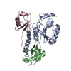
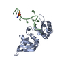
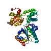
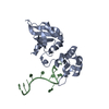
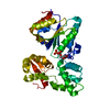
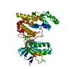
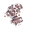
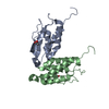
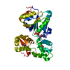
| Title | Three-dimensional structure of the protein XoxI (Q81AY6) from Bacillus cereus. Northeast Structural Genomics Consortium target BcR196. | ||||||
 Components Components | Protein XoxI | ||||||
 Keywords Keywords |  STRUCTURAL GENOMICS / UNKNOWN FUNCTION / Q81AY6 / NESG / XoxI / PSI-2 / Protein Structure Initiative / Northeast Structural Genomics Consortium STRUCTURAL GENOMICS / UNKNOWN FUNCTION / Q81AY6 / NESG / XoxI / PSI-2 / Protein Structure Initiative / Northeast Structural Genomics Consortium | ||||||
| Function / homology | Polyketide cyclase/dehydrase / Polyketide cyclase / dehydrase and lipid transport / START domain / Alpha-D-Glucose-1,6-Bisphosphate; Chain A, domain 4 / START-like domain superfamily / 2-Layer Sandwich / Alpha Beta / XoxI Function and homology information Function and homology information | ||||||
| Biological species |  Bacillus cereus ATCC 14579 (bacteria) Bacillus cereus ATCC 14579 (bacteria) | ||||||
| Method | X-RAY DIFFRACTION / SYNCHROTRON / SAD / Resolution: 2.48 Å | ||||||
 Authors Authors | Kuzin, A.P. / Abashidze, M. / Seetharaman, J. / Wang, H. / Ciccosanti, C. / Mao, L. / Xiao, R. / Nair, R. / Baran, M.C. / Acton, T.B. ...Kuzin, A.P. / Abashidze, M. / Seetharaman, J. / Wang, H. / Ciccosanti, C. / Mao, L. / Xiao, R. / Nair, R. / Baran, M.C. / Acton, T.B. / Rost, B. / Montelione, G.T. / Hunt, J.F. / Tong, L. / Northeast Structural Genomics Consortium (NESG) | ||||||
 Citation Citation | Journal: To Be Published Title: Three-dimensional structure of the protein XoxI (Q81AY6) from Bacillus cereus. Northeast Structural Genomics Consortium target BcR196. Authors: Kuzin, A.P. / Abashidze, M. / Seetharaman, J. / Wang, H. / Ciccosanti, C. / Mao, L. / Xiao, R. / Nair, R. / Baran, M.C. / Acton, T.B. / Rost, B. / Montelione, G.T. / Hunt, J.F. / Tong, L. | ||||||
| History |
|
- Structure visualization
| Structure viewer | Molecule: MolmilJmol/JSmol |
|---|
- Downloads & links
-Download
| PDBx/mmCIF format | 3cnw.cif.gz | 64.6 KB | Display | PDBx/mmCIF format |
|---|---|---|---|---|
| PDB format | pdb3cnw.ent.gz | 52.2 KB | Display | PDB format |
| PDBx/mmJSON format | 3cnw.json.gz | Tree view | PDBx/mmJSON format | |
| Others |  Other downloads Other downloads |
-Validation report
| Arichive directory | https://data.pdbj.org/pub/pdb/validation_reports/cn/3cnwftp://data.pdbj.org/pub/pdb/validation_reports/cn/3cnw | HTTPS FTP |
|---|
-Related structure data
| Similar structure data | |
|---|---|
| Other databases |

-Links
 PDBj
PDBj- Assembly
| Deposited unit | 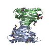
| ||||||||
|---|---|---|---|---|---|---|---|---|---|
| 1 |
| ||||||||
| 2 | 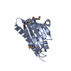
| ||||||||
| 3 | 
| ||||||||
| Unit cell |
|
-Components
| #1: Protein | Mass: 17039.254 Da / Num. of mol.: 2 Source method: isolated from a genetically manipulated source Source: (gene. exp.) Bacillus cereus ATCC 14579 (bacteria) / Species: Bacillus cereus / Strain: DSM 31 / Gene: BC_3411 / Production host: Escherichia coli (E. coli) / References: UniProt: Q81AY6#2: Water | ChemComp-HOH / | Water Mass: 18.015 Da / Num. of mol.: 60 / Source method: isolated from a natural source / Formula: H2O Mass: 18.015 Da / Num. of mol.: 60 / Source method: isolated from a natural source / Formula: H2O |
|---|
-Experimental details
-Experiment
| Experiment | Method: X-RAY DIFFRACTION / Number of used crystals: 1 |
|---|
- Sample preparation
| Crystal | Density Matthews: 1.93 Å3/Da / Density % sol: 36.32 % Description: The structure factor file contains Friedel pairs |
|---|---|
| Crystal grow | Temperature: 293 K / Method: vapor diffusion, hanging drop / pH: 4.6 Details: 0.1M Na Acetate, 30% PEG 550 MME, pH 4.6, VAPOR DIFFUSION, HANGING DROP, temperature 293K |
-Data collection
| Diffraction | Mean temperature: 100 K |
|---|---|
| Diffraction source | Source: SYNCHROTRON / Site: NSLS  / Beamline: X4A / Wavelength: 0.979 Å / Beamline: X4A / Wavelength: 0.979 Å |
| Detector | Type: ADSC QUANTUM 4 / Detector: CCD / Date: Feb 26, 2008 / Details: Mirrors |
| Radiation | Protocol: SINGLE WAVELENGTH / Monochromatic (M) / Laue (L): M / Scattering type: x-ray |
| Radiation wavelength | Wavelength: 0.979 Å / Relative weight: 1 |
| Reflection | Resolution: 2.48→50 Å / Num. obs: 17694 / % possible obs: 98.3 % / Observed criterion σ(I): -3 / Redundancy: 18 % / Biso Wilson estimate: 24.7 Å2 / Rmerge(I) obs: 0.097 / Net I/σ(I): 20 |
| Reflection shell | Resolution: 2.48→2.59 Å / Rmerge(I) obs: 0.19 / Mean I/σ(I) obs: 10.1 / % possible all: 84.3 |
- Processing
| Software |
| ||||||||||||||||||||
|---|---|---|---|---|---|---|---|---|---|---|---|---|---|---|---|---|---|---|---|---|---|
| Refinement | Method to determine structure: SAD / Resolution: 2.48→19.81 Å / Rfactor Rfree error: 0.009 / Data cutoff high absF: 97383.19 / Data cutoff low absF: 0 / Isotropic thermal model: RESTRAINED / Cross valid method: THROUGHOUT / σ(F): 1 / Stereochemistry target values: Engh & Huber Details: The Friedel pairs were used in phasing. BULK SOLVENT MODEL WAS USED IN REFINEMENT
| ||||||||||||||||||||
| Solvent computation | Solvent model: FLAT MODEL / Bsol: 51.1349 Å2 / ksol: 0.45 e/Å3 | ||||||||||||||||||||
| Displacement parameters | Biso mean: 28.6 Å2
| ||||||||||||||||||||
| Refine analyze |
| ||||||||||||||||||||
| Refinement step | Cycle: LAST / Resolution: 2.48→19.81 Å
| ||||||||||||||||||||
| Refine LS restraints |
| ||||||||||||||||||||
| LS refinement shell | Resolution: 2.48→2.55 Å / Rfactor Rfree error: 0.062 / Total num. of bins used: 6
| ||||||||||||||||||||
| Xplor file |
|
