 Movie
Movie Controller

[English] 日本語
 Yorodumi
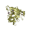
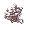
- PDB-3cnm: Crystal Structure of Phenazine Biosynthesis Protein PhzA/B from B... -
+ Open data

- Basic information
| Entry | Database: PDB / ID: 3cnm | ||||||
|---|---|---|---|---|---|---|---|
| Title | Crystal Structure of Phenazine Biosynthesis Protein PhzA/B from Burkholderia cepacia R18194, DHHA complex | ||||||
 Components Components | Phenazine biosynthesis protein A/B | ||||||
 Keywords Keywords |  BIOSYNTHETIC PROTEIN / phenazine biosynthesis / imine / Schiff base BIOSYNTHETIC PROTEIN / phenazine biosynthesis / imine / Schiff base | ||||||
| Function / homology |  Function and homology information Function and homology information | ||||||
| Biological species |  Burkholderia sp. (bacteria) Burkholderia sp. (bacteria) | ||||||
| Method | X-RAY DIFFRACTION / SYNCHROTRON / rigid body refinement of apo structure / Resolution: 1.65 Å | ||||||
 Authors Authors | Ahuja, E.G. / Blankenfeldt, W. | ||||||
 Citation Citation | Journal: J.Am.Chem.Soc. / Year: 2008 Title: PhzA/B catalyzes the formation of the tricycle in phenazine biosynthesis. Authors: Ahuja, E.G. / Janning, P. / Mentel, M. / Graebsch, A. / Breinbauer, R. / Hiller, W. / Costisella, B. / Thomashow, L.S. / Mavrodi, D.V. / Blankenfeldt, W. | ||||||
| History |
|
- Structure visualization
| Structure viewer | Molecule: MolmilJmol/JSmol |
|---|
- Downloads & links
-Download
| PDBx/mmCIF format | 3cnm.cif.gz | 92.5 KB | Display | PDBx/mmCIF format |
|---|---|---|---|---|
| PDB format | pdb3cnm.ent.gz | 69.6 KB | Display | PDB format |
| PDBx/mmJSON format | 3cnm.json.gz | Tree view | PDBx/mmJSON format | |
| Others |  Other downloads Other downloads |
-Validation report
| Arichive directory | https://data.pdbj.org/pub/pdb/validation_reports/cn/3cnmftp://data.pdbj.org/pub/pdb/validation_reports/cn/3cnm | HTTPS FTP |
|---|
-Related structure data
| Related structure data | 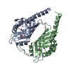 3b4oSC  3b4pC 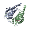 3dzlC  3ex9C S: Starting model for refinement C: citing same article ( |
|---|---|
| Similar structure data |


-Links
 PDBj
PDBj
- Assembly
| Deposited unit | 
| ||||||||
|---|---|---|---|---|---|---|---|---|---|
| 1 |
| ||||||||
| Unit cell |
| ||||||||
| Details | dimer |
-Components
| #1: Protein | Mass: 21531.025 Da / Num. of mol.: 2 Source method: isolated from a genetically manipulated source Source: (gene. exp.) Burkholderia sp. (bacteria) / Strain: R18194 / Plasmid: pET15b / Production host: Escherichia coli (E. coli) / Strain (production host): Rosetta pLysS / References: UniProt: Q396C9#2: Chemical | Acetate  Mass: 59.044 Da / Num. of mol.: 3 / Source method: obtained synthetically / Formula: C2H3O2 Mass: 59.044 Da / Num. of mol.: 3 / Source method: obtained synthetically / Formula: C2H3O2#3: Chemical |   Mass: 155.151 Da / Num. of mol.: 2 / Source method: obtained synthetically / Formula: C7H9NO3 Mass: 155.151 Da / Num. of mol.: 2 / Source method: obtained synthetically / Formula: C7H9NO3#4: Water | ChemComp-HOH / | Water Mass: 18.015 Da / Num. of mol.: 377 / Source method: isolated from a natural source / Formula: H2O Mass: 18.015 Da / Num. of mol.: 377 / Source method: isolated from a natural source / Formula: H2O |
|---|
-Experimental details
-Experiment
| Experiment | Method: X-RAY DIFFRACTION / Number of used crystals: 1 |
|---|
- Sample preparation
| Crystal | Density Matthews: 2.29 Å3/Da / Density % sol: 46.38 % |
|---|---|
| Crystal grow | Temperature: 285 K / Method: vapor diffusion, hanging drop / pH: 6.1 Details: 16-20% (w/v) PEG3350, 0.2 M NH4OAc, 0.1 M Bis-Tris pH 6.1-6.7; complex prepared by overnight soaking in mother liquor containing 50 mM DHHA, VAPOR DIFFUSION, HANGING DROP, temperature 285K |
-Data collection
| Diffraction | Mean temperature: 100 K |
|---|---|
| Diffraction source | Source: SYNCHROTRON / Site: SLS  / Beamline: X10SA / Wavelength: 0.9809 Å / Beamline: X10SA / Wavelength: 0.9809 Å |
| Detector | Type: MARMOSAIC 225 mm CCD / Detector: CCD / Date: Mar 6, 2008 / Details: SI(111) monochromator |
| Radiation | Monochromator: SI(111) / Protocol: SINGLE WAVELENGTH / Monochromatic (M) / Laue (L): M / Scattering type: x-ray |
| Radiation wavelength | Wavelength: 0.9809 Å / Relative weight: 1 |
| Reflection | Resolution: 1.65→20 Å / Num. all: 48683 / Num. obs: 48675 / % possible obs: 100 % / Observed criterion σ(I): 3 / Redundancy: 14.1 % / Biso Wilson estimate: 33 Å2 / Rmerge(I) obs: 0.066 / Rsym value: 0.139 / Net I/σ(I): 16 |
| Reflection shell | Resolution: 1.65→1.75 Å / Redundancy: 13.3 % / Rmerge(I) obs: 0.42 / Mean I/σ(I) obs: 3.7 / Num. unique all: 7754 / % possible all: 100 |
- Processing
| Software |
| |||||||||||||||||||||||||||||||||||||||||||||||||||||||||||||||||||||||||||||||||||||||||||||||||||||||||||||||||||||||||||||
|---|---|---|---|---|---|---|---|---|---|---|---|---|---|---|---|---|---|---|---|---|---|---|---|---|---|---|---|---|---|---|---|---|---|---|---|---|---|---|---|---|---|---|---|---|---|---|---|---|---|---|---|---|---|---|---|---|---|---|---|---|---|---|---|---|---|---|---|---|---|---|---|---|---|---|---|---|---|---|---|---|---|---|---|---|---|---|---|---|---|---|---|---|---|---|---|---|---|---|---|---|---|---|---|---|---|---|---|---|---|---|---|---|---|---|---|---|---|---|---|---|---|---|---|---|---|---|
| Refinement | Method to determine structure: rigid body refinement of apo structure Starting model: 3B4O Resolution: 1.65→19.81 Å / Cor.coef. Fo:Fc: 0.972 / Cor.coef. Fo:Fc free: 0.958 / SU B: 3.23 / SU ML: 0.051 / TLS residual ADP flag: LIKELY RESIDUAL / Cross valid method: THROUGHOUT / ESU R: 0.079 / ESU R Free: 0.085 / Stereochemistry target values: MAXIMUM LIKELIHOOD Details: HYDROGENS HAVE BEEN ADDED IN THE RIDING POSITIONS; TLS-Refinement was used throughout (1 TLS body)
| |||||||||||||||||||||||||||||||||||||||||||||||||||||||||||||||||||||||||||||||||||||||||||||||||||||||||||||||||||||||||||||
| Solvent computation | Ion probe radii: 0.8 Å / Shrinkage radii: 0.8 Å / VDW probe radii: 1.2 Å / Solvent model: BABINET MODEL WITH MASK | |||||||||||||||||||||||||||||||||||||||||||||||||||||||||||||||||||||||||||||||||||||||||||||||||||||||||||||||||||||||||||||
| Displacement parameters | Biso mean: 24.467 Å2 | |||||||||||||||||||||||||||||||||||||||||||||||||||||||||||||||||||||||||||||||||||||||||||||||||||||||||||||||||||||||||||||
| Refinement step | Cycle: LAST / Resolution: 1.65→19.81 Å
| |||||||||||||||||||||||||||||||||||||||||||||||||||||||||||||||||||||||||||||||||||||||||||||||||||||||||||||||||||||||||||||
| Refine LS restraints |
| |||||||||||||||||||||||||||||||||||||||||||||||||||||||||||||||||||||||||||||||||||||||||||||||||||||||||||||||||||||||||||||
| LS refinement shell | Resolution: 1.65→1.693 Å / Total num. of bins used: 20
| |||||||||||||||||||||||||||||||||||||||||||||||||||||||||||||||||||||||||||||||||||||||||||||||||||||||||||||||||||||||||||||
| Refinement TLS params. | Method: refined / Origin x: 37.0061 Å / Origin y: 5.9718 Å / Origin z: 79.8364 Å
| |||||||||||||||||||||||||||||||||||||||||||||||||||||||||||||||||||||||||||||||||||||||||||||||||||||||||||||||||||||||||||||
| Refinement TLS group |
|
