DNA polymerase V complex / homologous recombination / recombinational repair / SOS response / ATP-dependent DNA damage sensor activity / response to ionizing radiation / translesion synthesis / ATP-dependent activity, acting on DNA / cell motility / single-stranded DNA binding ...DNA polymerase V complex / homologous recombination / recombinational repair / SOS response / ATP-dependent DNA damage sensor activity / response to ionizing radiation / translesion synthesis / ATP-dependent activity, acting on DNA / cell motility / single-stranded DNA binding / DNA-binding transcription factor binding / DNA recombination / damaged DNA binding / DNA damage response / ATP hydrolysis activity / ATP binding / cytoplasm Similarity search - Function
RecA protein, C-terminal domain / Rec A Protein; domain 2 / : / : / RecA C-terminal domain / DNA recombination/repair protein RecA, conserved site / DNA recombination and repair protein RecA, C-terminal / recA signature. / DNA recombination and repair protein RecA / recA bacterial DNA recombination protein ...RecA protein, C-terminal domain / Rec A Protein; domain 2 / : / : / RecA C-terminal domain / DNA recombination/repair protein RecA, conserved site / DNA recombination and repair protein RecA, C-terminal / recA signature. / DNA recombination and repair protein RecA / recA bacterial DNA recombination protein / DNA recombination and repair protein RecA, monomer-monomer interface / RecA family profile 2. / DNA recombination and repair protein RecA-like, ATP-binding domain / RecA family profile 1. / P-loop containing nucleotide triphosphate hydrolases / ATPases associated with a variety of cellular activities / AAA+ ATPase domain / P-loop containing nucleoside triphosphate hydrolase / Rossmann fold / 2-Layer Sandwich / 3-Layer(aba) Sandwich / Alpha Beta Similarity search - Domain/homology
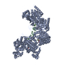
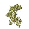
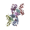
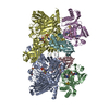
B: DNA (5'-D(*DTP*DTP*DTP*DTP*DTP*DTP*DTP*DTP*DTP*DTP*DTP*DTP*DTP*DTP*DT)-3') C: DNA (5'-D(*DAP*DAP*DAP*DAP*DAP*DAP*DAP*DAP*DAP*DAP*DAP*DA)-3') E: DNA (5'-D(*DTP*DTP*DTP*DTP*DTP*DTP*DTP*DTP*DTP*DTP*DTP*DTP*DTP*DTP*DT)-3') F: DNA (5'-D(*DAP*DAP*DAP*DAP*DAP*DAP*DAP*DAP*DAP*DAP*DAP*DA)-3') A: Protein recA D: Protein recA hetero molecules
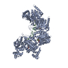
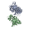
B: DNA (5'-D(*DTP*DTP*DTP*DTP*DTP*DTP*DTP*DTP*DTP*DTP*DTP*DTP*DTP*DTP*DT)-3') C: DNA (5'-D(*DAP*DAP*DAP*DAP*DAP*DAP*DAP*DAP*DAP*DAP*DAP*DA)-3') A: Protein recA hetero molecules
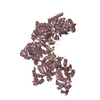
E: DNA (5'-D(*DTP*DTP*DTP*DTP*DTP*DTP*DTP*DTP*DTP*DTP*DTP*DTP*DTP*DTP*DT)-3') F: DNA (5'-D(*DAP*DAP*DAP*DAP*DAP*DAP*DAP*DAP*DAP*DAP*DAP*DA)-3') D: Protein recA hetero molecules
Density Matthews: 2.51 Å3/Da / Density % sol: 51.03 %
Crystal grow
pH: 8 Details: RecA5 fusion protein was incubated with a 3-fold molar excess of ssDNA in the original protein buffer supplemented with 2 mM ADP, 10 mM MgCl2 and 8 mM AlF4, pH 6.0. Crystals of the RecA5- ...Details: RecA5 fusion protein was incubated with a 3-fold molar excess of ssDNA in the original protein buffer supplemented with 2 mM ADP, 10 mM MgCl2 and 8 mM AlF4, pH 6.0. Crystals of the RecA5-(ADP-AlF4-Mg)5-(dT)15 complex were grown from 50 mM Tris-Cl, 9% (w/v) PVP K15, 32% (v/v) MPD, 10 mM DTT, pH 8.0. RecA5-(ADP-AlF4-Mg)5-(dT)15-(dA)12 complex were obtained by soaking the RecA5-(ADP-AlF4-Mg)5-(dT)15 crystals in a 0.2 mM solution of the complementary (dA)12 oligonucleotide in 25 mM Tris-Cl, 9% (w/v) PVP K15, 32% (v/v) MPD, 2 mM ADP, 8 mM AlF4, and 10 mM MgCl2 for 4 hr.
In the structure databanks used in Yorodumi, some data are registered as the other names, "COVID-19 virus" and "2019-nCoV". Here are the details of the virus and the list of structure data.
Jan 31, 2019. EMDB accession codes are about to change! (news from PDBe EMDB page)
EMDB accession codes are about to change! (news from PDBe EMDB page)
The allocation of 4 digits for EMDB accession codes will soon come to an end. Whilst these codes will remain in use, new EMDB accession codes will include an additional digit and will expand incrementally as the available range of codes is exhausted. The current 4-digit format prefixed with “EMD-” (i.e. EMD-XXXX) will advance to a 5-digit format (i.e. EMD-XXXXX), and so on. It is currently estimated that the 4-digit codes will be depleted around Spring 2019, at which point the 5-digit format will come into force.
The EM Navigator/Yorodumi systems omit the EMD- prefix.
Related info.:Q: What is EMD? / ID/Accession-code notation in Yorodumi/EM Navigator
Yorodumi is a browser for structure data from EMDB, PDB, SASBDB, etc.
This page is also the successor to EM Navigator detail page, and also detail information page/front-end page for Omokage search.
The word "yorodu" (or yorozu) is an old Japanese word meaning "ten thousand". "mi" (miru) is to see.
Related info.:EMDB / PDB / SASBDB / Comparison of 3 databanks / Yorodumi Search / Aug 31, 2016. New EM Navigator & Yorodumi / Yorodumi Papers / Jmol/JSmol / Function and homology information / Changes in new EM Navigator and Yorodumi
 Movie
Movie Controller

 Yorodumi
 Open data
 Basic information
 Components
 Keywords
 homologous recombination / recombination-DNA COMPLEX
 Function and homology information
 Authors
 Citation
 Structure visualization
 Downloads & links
 Other downloads


 PDBj
PDBj


 Assembly
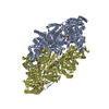


 Mass: 24.305 Da / Num. of mol.: 10 / Source method: obtained synthetically / Formula: Mg
 Mass: 102.975 Da / Num. of mol.: 10 / Source method: obtained synthetically / Formula: AlF4
 Mass: 427.201 Da / Num. of mol.: 10 / Source method: obtained synthetically / Formula: C10H15N5O10P2 / Comment: ADP, energy-carrying molecule*YM
 Sample preparation
 / Beamline: 24-ID-C
 Processing