 Movie
Movie Controller

[English] 日本語
 Yorodumi
- PDB-3cms: ENGINEERING ENZYME SUB-SITE SPECIFICITY: PREPARATION, KINETIC CHA... -
+ Open data

- Basic information
| Entry | Database: PDB / ID: 3cms | ||||||
|---|---|---|---|---|---|---|---|
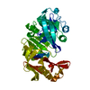
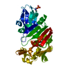
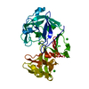
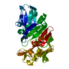
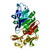
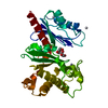
| Title | ENGINEERING ENZYME SUB-SITE SPECIFICITY: PREPARATION, KINETIC CHARACTERIZATION AND X-RAY ANALYSIS AT 2.0-ANGSTROMS RESOLUTION OF VAL111PHE SITE-MUTATED CALF CHYMOSIN | ||||||
 Components Components | CHYMOSIN B | ||||||
 Keywords Keywords |  HYDROLASE / ACID PROTEINASE HYDROLASE / ACID PROTEINASE | ||||||
| Function / homology |  Function and homology informationchymosin / digestion / aspartic-type endopeptidase activity / proteolysis Function and homology informationchymosin / digestion / aspartic-type endopeptidase activity / proteolysisSimilarity search - Function | ||||||
| Biological species |  Bos taurus (cattle) Bos taurus (cattle) | ||||||
| Method | X-RAY DIFFRACTION / Resolution: 2 Å | ||||||
 Authors Authors | Newman, M. / Frazao, C. / Shearer, A. / Tickle, I.J. / Blundell, T.L. | ||||||
 Citation Citation | Journal: Biochemistry / Year: 1990 Title: Engineering enzyme subsite specificity: preparation, kinetic characterization, and X-ray analysis at 2.0-A resolution of Val111Phe site-mutated calf chymosin. Authors: Strop, P. / Sedlacek, J. / Stys, J. / Kaderabkova, Z. / Blaha, I. / Pavlickova, L. / Pohl, J. / Fabry, M. / Kostka, V. / Newman, M. #1: Journal: J.Mol.Biol. / Year: 1991Title: X-Ray Analyses of Aspartic Proteinases Iv: Structure and Refinement at 2.2 Angstroms Resolution of Bovine Chymosin Authors: Newman, M. / Safro, M. / Frazao, C. / Kahn, G. / Zdanov, A. / Tickle, I.J. / Blundell, T.L. / Andreeva, N. | ||||||
| History |
|
- Structure visualization
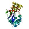
| Structure viewer | Molecule: MolmilJmol/JSmol |
|---|
- Downloads & links
-Download
| PDBx/mmCIF format | 3cms.cif.gz | 73.1 KB | Display | PDBx/mmCIF format |
|---|---|---|---|---|
| PDB format | pdb3cms.ent.gz | 56.8 KB | Display | PDB format |
| PDBx/mmJSON format | 3cms.json.gz | Tree view | PDBx/mmJSON format | |
| Others |  Other downloads Other downloads |
-Validation report
| Arichive directory | https://data.pdbj.org/pub/pdb/validation_reports/cm/3cmsftp://data.pdbj.org/pub/pdb/validation_reports/cm/3cms | HTTPS FTP |
|---|
-Related structure data
| Similar structure data |
|---|

-Links
 PDBj
PDBj
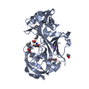
- Assembly
| Deposited unit | 
| ||||||||
|---|---|---|---|---|---|---|---|---|---|
| 1 |
| ||||||||
| Unit cell |
| ||||||||
| Atom site foot note | 1: RESIDUE PRO 23 IS A CIS-PROLINE. 2: THESE RESIDUES ARE IN POORLY DEFINED REGIONS IN THE ELECTRON DENSITY MAP. 3: RESIDUES 72 TO 79 HAVE BEEN REFINED IN TWO ALTERNATE CONFORMATIONS. THESE RESIDUES ARE IN POORLY DEFINED REGIONS IN THE ELECTRON DENSITY MAP. CONFORMATION B HAS THE HIGHER RELATIVE OCCUPANCY (O.60) ...3: RESIDUES 72 TO 79 HAVE BEEN REFINED IN TWO ALTERNATE CONFORMATIONS. THESE RESIDUES ARE IN POORLY DEFINED REGIONS IN THE ELECTRON DENSITY MAP. CONFORMATION B HAS THE HIGHER RELATIVE OCCUPANCY (O.60) AND IS ASSOCIATED WITH SUPERIOR ELECTRON DENSITY. THUS IT APPEARS THAT CONFORMATION B IS SIGNIFICANTLY MORE HIGHLY POPULATED THAN CONFORMATION A. CONFORMATION A WITH OCCUPANCY OF 0.40 MUST BE REGARDED AS A TENTATIVE INTERPRETATION. |
-Components
| #1: Protein | Mass: 35720.738 Da / Num. of mol.: 1 Source method: isolated from a genetically manipulated source Source: (gene. exp.) Bos taurus (cattle) / References: UniProt: P00794, chymosin |
|---|---|
| #2: Water | ChemComp-HOH / Water Mass: 18.015 Da / Num. of mol.: 145 / Source method: isolated from a natural source / Formula: H2O Mass: 18.015 Da / Num. of mol.: 145 / Source method: isolated from a natural source / Formula: H2O |
| Compound details | THE SPECIFIC GENE MUTATION V111F IS SITUATED BETWEEN THE PRIMARY SPECIFICITY BINDING POCKET S1 AND ...THE SPECIFIC GENE MUTATION V111F IS SITUATED BETWEEN THE PRIMARY SPECIFICIT |
-Experimental details
-Experiment
| Experiment | Method: X-RAY DIFFRACTION |
|---|
- Sample preparation
| Crystal | Density Matthews: 2.33 Å3/Da / Density % sol: 47.16 % | ||||||||||||||||||||||||
|---|---|---|---|---|---|---|---|---|---|---|---|---|---|---|---|---|---|---|---|---|---|---|---|---|---|
| Crystal grow | *PLUS pH: 6.5 / Method: microdialysis | ||||||||||||||||||||||||
| Components of the solutions | *PLUS
|
-Data collection
| Radiation | Scattering type: x-ray |
|---|---|
| Radiation wavelength | Relative weight: 1 |
| Reflection | *PLUS Highest resolution: 2 Å / Num. obs: 22098 / % possible obs: 96.4 % / Observed criterion σ(I): 0.072 / Num. measured all: 149939 / Rmerge(I) obs: 3 |
- Processing
| Software | Name: RESTRAIN / Classification: refinement | |||||||||||||||||||||||||||||||||||||||||||||||||||||||||||||||
|---|---|---|---|---|---|---|---|---|---|---|---|---|---|---|---|---|---|---|---|---|---|---|---|---|---|---|---|---|---|---|---|---|---|---|---|---|---|---|---|---|---|---|---|---|---|---|---|---|---|---|---|---|---|---|---|---|---|---|---|---|---|---|---|---|
| Refinement | Resolution: 2→10 Å Details: THE QUANTITY PRESENTED IN THE TEMPERATURE FACTOR FIELD IS U. RESIDUES 72 TO 79 HAVE BEEN REFINED IN TWO ALTERNATE CONFORMATIONS. TURN 5 WITH THE A CONFORMATION IS CLASSIFED TYPE II' 2:2 ...Details: THE QUANTITY PRESENTED IN THE TEMPERATURE FACTOR FIELD IS U. RESIDUES 72 TO 79 HAVE BEEN REFINED IN TWO ALTERNATE CONFORMATIONS. TURN 5 WITH THE A CONFORMATION IS CLASSIFED TYPE II' 2:2 DISTORTED. TURN 5 WITH THE B CONFORMATION IS UNCLASSIFIED 2:2. INVARIANT RESIDUE TYR 14 HAS BEEN BUILT INTO A CONFORMATION THAT DIFFERS FROM THE WILD-TYPE CHYMOSIN STRUCTURE. HOWEVER, THIS RESIDUE BELONGS TO THE POORLY DEFINED SURFACE REGION BETWEEN STRANDS AN AND BN, AND THUS THE ASSIGNMENT OF ITS POSITION MUST BE REGARDED AS TENTATIVE.
| |||||||||||||||||||||||||||||||||||||||||||||||||||||||||||||||
| Refinement step | Cycle: LAST / Resolution: 2→10 Å
| |||||||||||||||||||||||||||||||||||||||||||||||||||||||||||||||
| Refine LS restraints |
| |||||||||||||||||||||||||||||||||||||||||||||||||||||||||||||||
| Refinement | *PLUS Rfactor obs: 0.195 | |||||||||||||||||||||||||||||||||||||||||||||||||||||||||||||||
| Solvent computation | *PLUS | |||||||||||||||||||||||||||||||||||||||||||||||||||||||||||||||
| Displacement parameters | *PLUS Biso mean: 30.3 Å2 | |||||||||||||||||||||||||||||||||||||||||||||||||||||||||||||||
| Refine LS restraints | *PLUS Type: p_plane_restr / Dev ideal: 0.019 |
