 Movie
Movie Controller


+ Open data

- Basic information
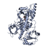
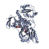
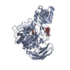
| Entry | Database: PDB / ID: 3cm8 | ||||||
|---|---|---|---|---|---|---|---|
| Title | A RNA polymerase subunit structure from virus | ||||||
 Components Components |
| ||||||
 Keywords Keywords | RNA BINDING PROTEIN/Transferase / Protein-peptide Complex / Nucleotide-binding /  Nucleotidyltransferase / RNA replication / RNA-directed RNA polymerase / Transferase / RNA BINDING PROTEIN-Transferase COMPLEX Nucleotidyltransferase / RNA replication / RNA-directed RNA polymerase / Transferase / RNA BINDING PROTEIN-Transferase COMPLEX | ||||||
| Function / homology |  Function and homology information Function and homology informationsymbiont-mediated suppression of host mRNA transcription via inhibition of RNA polymerase II activity / cap snatching / viral transcription / endonuclease activity / host cell cytoplasm / Hydrolases; Acting on ester bonds / RNA-directed RNA polymerase / viral RNA genome replication / RNA-dependent RNA polymerase activity / nucleotide binding ...symbiont-mediated suppression of host mRNA transcription via inhibition of RNA polymerase II activity / cap snatching / viral transcription / endonuclease activity / host cell cytoplasm / Hydrolases; Acting on ester bonds / RNA-directed RNA polymerase / viral RNA genome replication / RNA-dependent RNA polymerase activity / nucleotide binding / DNA-templated transcription / host cell nucleus / RNA binding / metal ion bindingSimilarity search - Function | ||||||
| Biological species |   Influenza A virus Influenza A virus | ||||||
| Method | X-RAY DIFFRACTION / SYNCHROTRON / MAD / Resolution: 2.899 Å | ||||||
 Authors Authors | He, X. / Zhou, J. / Zeng, Z. / Ma, J. / Zhang, R. / Rao, Z. / Liu, Y. | ||||||
 Citation Citation | Journal: Nature / Year: 2008 Title: Crystal structure of the polymerase PAC-PB1N complex from an avian influenza H5N1 virus Authors: He, X. / Zhou, J. / Bartlam, M. / Zhang, R. / Ma, J. / Lou, Z. / Li, X. / Li, J. / Joachimiak, A. / Zeng, Z. / Ge, R. / Rao, Z. / Liu, Y. | ||||||
| History |
|
- Structure visualization
| Structure viewer | Molecule: MolmilJmol/JSmol |
|---|
- Downloads & links
-Download
| PDBx/mmCIF format | 3cm8.cif.gz | 103.6 KB | Display | PDBx/mmCIF format |
|---|---|---|---|---|
| PDB format | pdb3cm8.ent.gz | 79.6 KB | Display | PDB format |
| PDBx/mmJSON format | 3cm8.json.gz | Tree view | PDBx/mmJSON format | |
| Others |  Other downloads Other downloads |
-Validation report
| Arichive directory | https://data.pdbj.org/pub/pdb/validation_reports/cm/3cm8ftp://data.pdbj.org/pub/pdb/validation_reports/cm/3cm8 | HTTPS FTP |
|---|
-Related structure data
| Similar structure data |
|---|


-Links
 PDBj
PDBj- Assembly
| Deposited unit | 
| ||||||||
|---|---|---|---|---|---|---|---|---|---|
| 1 |
| ||||||||
| Unit cell |
| ||||||||
| Components on special symmetry positions |
|
-Components
| #1: Protein | Mass: 53870.789 Da / Num. of mol.: 1 / Fragment: residues in database 256-716 Source method: isolated from a genetically manipulated source Source: (gene. exp.) Influenza A virus / Strain: A/Environment/Hong Kong/437-6/99 / Production host:  Escherichia coli (E. coli) / References: UniProt: Q9EA60 Escherichia coli (E. coli) / References: UniProt: Q9EA60 |
|---|---|
| #2: Protein/peptide | / Polymerase basic protein 1 / PB1 / RNA-directed RNA polymerase subunit P1 Mass: 3194.674 Da / Num. of mol.: 1 Source method: isolated from a genetically manipulated source Source: (gene. exp.) Influenza A virus / Strain: A/Hong Kong/156/1997 H5N1 genotype Gs/Gd / Production host: Escherichia coli (E. coli) / References: UniProt: Q9WLS3, RNA-directed RNA polymerase |
| #3: Water | ChemComp-HOH / Water Mass: 18.015 Da / Num. of mol.: 49 / Source method: isolated from a natural source / Formula: H2O Mass: 18.015 Da / Num. of mol.: 49 / Source method: isolated from a natural source / Formula: H2O |
-Experimental details
-Experiment
| Experiment | Method: X-RAY DIFFRACTION / Number of used crystals: 2 |
|---|
- Sample preparation
| Crystal | Density Matthews: 4.38 Å3/Da / Density % sol: 71.94 % |
|---|---|
| Crystal grow | Temperature: 289 K / Method: vapor diffusion, hanging drop / pH: 8 Details: 1M sodium acetate, pH 8.0, VAPOR DIFFUSION, HANGING DROP, temperature 289K |
-Data collection
| Diffraction |
| ||||||||||||||||||
|---|---|---|---|---|---|---|---|---|---|---|---|---|---|---|---|---|---|---|---|
| Diffraction source |
| ||||||||||||||||||
| Detector |
| ||||||||||||||||||
| Radiation |
| ||||||||||||||||||
| Radiation wavelength |
| ||||||||||||||||||
| Reflection | Resolution: 2.899→50 Å / Num. all: 23042 / Num. obs: 22964 / % possible obs: 99.7 % / Observed criterion σ(F): 4.6 / Observed criterion σ(I): 4.6 / Biso Wilson estimate: 75.44 Å2 | ||||||||||||||||||
| Reflection shell | Highest resolution: 2.899 Å / % possible all: 99 |

- Processing
| Software |
| |||||||||||||||||||||||||||||||||||||||||||||||||||||||||||||||
|---|---|---|---|---|---|---|---|---|---|---|---|---|---|---|---|---|---|---|---|---|---|---|---|---|---|---|---|---|---|---|---|---|---|---|---|---|---|---|---|---|---|---|---|---|---|---|---|---|---|---|---|---|---|---|---|---|---|---|---|---|---|---|---|---|
| Refinement | Method to determine structure: MAD / Resolution: 2.899→45.184 Å / FOM work R set: 0.787 / σ(F): 1.31 / Stereochemistry target values: ML
| |||||||||||||||||||||||||||||||||||||||||||||||||||||||||||||||
| Solvent computation | Shrinkage radii: 0.9 Å / VDW probe radii: 1.11 Å / Solvent model: FLAT BULK SOLVENT MODEL / Bsol: 80.261 Å2 / ksol: 0.371 e/Å3 | |||||||||||||||||||||||||||||||||||||||||||||||||||||||||||||||
| Displacement parameters | Biso max: 42.23 Å2 / Biso mean: 84.28 Å2 / Biso min: 166.55 Å2
| |||||||||||||||||||||||||||||||||||||||||||||||||||||||||||||||
| Refinement step | Cycle: LAST / Resolution: 2.899→45.184 Å
| |||||||||||||||||||||||||||||||||||||||||||||||||||||||||||||||
| Refine LS restraints |
| |||||||||||||||||||||||||||||||||||||||||||||||||||||||||||||||
| LS refinement shell | Refine-ID: X-RAY DIFFRACTION / Total num. of bins used: 8
|
