 Movie
Movie Controller


+ Open data

- Basic information
| Entry | Database: PDB / ID: 3ckr | ||||||
|---|---|---|---|---|---|---|---|
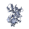
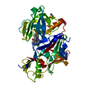
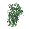
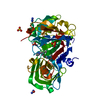
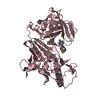
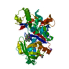
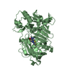
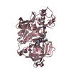
| Title | Crystal structure of BACE-1 in complex with inhibitor | ||||||
 Components Components | Beta-secretase 1 | ||||||
 Keywords Keywords | HYDROLASE / beta-secretase / aspartyl protease / Alternative splicing / Glycoprotein / Membrane / Transmembrane / Zymogen | ||||||
| Function / homology |  Function and homology informationmemapsin 2 / Golgi-associated vesicle lumen / signaling receptor ligand precursor processing / beta-aspartyl-peptidase activity / amyloid precursor protein catabolic process / amyloid-beta formation / membrane protein ectodomain proteolysis / cellular response to manganese ion / amyloid-beta metabolic process / prepulse inhibition ...memapsin 2 / Golgi-associated vesicle lumen / signaling receptor ligand precursor processing / beta-aspartyl-peptidase activity / amyloid precursor protein catabolic process / amyloid-beta formation / membrane protein ectodomain proteolysis / cellular response to manganese ion / amyloid-beta metabolic process / prepulse inhibition / detection of mechanical stimulus involved in sensory perception of pain / cellular response to copper ion / presynaptic modulation of chemical synaptic transmission / hippocampal mossy fiber to CA3 synapse / multivesicular body / response to lead ion / trans-Golgi network / recycling endosome / protein processing / cellular response to amyloid-beta / positive regulation of neuron apoptotic process / synaptic vesicle / late endosome / peptidase activity / amyloid-beta binding / endopeptidase activity / amyloid fibril formation / lysosome / aspartic-type endopeptidase activity / early endosome / endosome membrane / endosome / membrane raft / Amyloid fiber formation / endoplasmic reticulum lumen / axon / neuronal cell body / dendrite / Golgi apparatus / enzyme binding / cell surface / proteolysis / membrane / plasma membrane Function and homology informationmemapsin 2 / Golgi-associated vesicle lumen / signaling receptor ligand precursor processing / beta-aspartyl-peptidase activity / amyloid precursor protein catabolic process / amyloid-beta formation / membrane protein ectodomain proteolysis / cellular response to manganese ion / amyloid-beta metabolic process / prepulse inhibition ...memapsin 2 / Golgi-associated vesicle lumen / signaling receptor ligand precursor processing / beta-aspartyl-peptidase activity / amyloid precursor protein catabolic process / amyloid-beta formation / membrane protein ectodomain proteolysis / cellular response to manganese ion / amyloid-beta metabolic process / prepulse inhibition / detection of mechanical stimulus involved in sensory perception of pain / cellular response to copper ion / presynaptic modulation of chemical synaptic transmission / hippocampal mossy fiber to CA3 synapse / multivesicular body / response to lead ion / trans-Golgi network / recycling endosome / protein processing / cellular response to amyloid-beta / positive regulation of neuron apoptotic process / synaptic vesicle / late endosome / peptidase activity / amyloid-beta binding / endopeptidase activity / amyloid fibril formation / lysosome / aspartic-type endopeptidase activity / early endosome / endosome membrane / endosome / membrane raft / Amyloid fiber formation / endoplasmic reticulum lumen / axon / neuronal cell body / dendrite / Golgi apparatus / enzyme binding / cell surface / proteolysis / membrane / plasma membraneSimilarity search - Function | ||||||
| Biological species |  Homo sapiens (human) Homo sapiens (human) | ||||||
| Method | X-RAY DIFFRACTION / SYNCHROTRON / MOLECULAR REPLACEMENT / molecular replacement / Resolution: 2.7 Å | ||||||
 Authors Authors | Min, K. | ||||||
 Citation Citation | Journal: Bioorg.Med.Chem.Lett. / Year: 2008 Title: Synthesis, SAR, and X-ray structure of human BACE-1 inhibitors with cyclic urea derivatives Authors: Park, H. / Min, K. / Kwak, H.-S. / Koo, K.D. / Lim, D. / Seo, S.-W. / Choi, J.-U. / Platt, B. / Choi, D.-Y. | ||||||
| History |
|
- Structure visualization
| Structure viewer | Molecule: MolmilJmol/JSmol |
|---|
- Downloads & links
-Download
| PDBx/mmCIF format | 3ckr.cif.gz | 230.8 KB | Display | PDBx/mmCIF format |
|---|---|---|---|---|
| PDB format | pdb3ckr.ent.gz | 193 KB | Display | PDB format |
| PDBx/mmJSON format | 3ckr.json.gz | Tree view | PDBx/mmJSON format | |
| Others |  Other downloads Other downloads |
-Validation report
| Arichive directory | https://data.pdbj.org/pub/pdb/validation_reports/ck/3ckrftp://data.pdbj.org/pub/pdb/validation_reports/ck/3ckr | HTTPS FTP |
|---|
-Related structure data


-Links
 PDBj
PDBj

- Assembly
| Deposited unit | 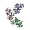
| ||||||||
|---|---|---|---|---|---|---|---|---|---|
| 1 | 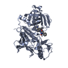
| ||||||||
| 2 | 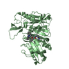
| ||||||||
| 3 | 
| ||||||||
| Unit cell |
|
-Components
| #1: Protein | / Beta-site APP cleaving enzyme 1 / Beta-site amyloid precursor protein cleaving enzyme 1 / Membrane- ...Beta-site APP cleaving enzyme 1 / Beta-site amyloid precursor protein cleaving enzyme 1 / Membrane-associated aspartic protease 2 / Memapsin-2 / Aspartyl protease 2 / Asp 2 / ASP2 Mass: 45923.547 Da / Num. of mol.: 3 / Fragment: protease domain, UNP residues 43-454 / Mutation: R-6K, R-5K Source method: isolated from a genetically manipulated source Source: (gene. exp.) Homo sapiens (human) / Production host:  Escherichia coli (E. coli) / References: UniProt: P56817, memapsin 2 Escherichia coli (E. coli) / References: UniProt: P56817, memapsin 2#2: Chemical |   Mass: 605.769 Da / Num. of mol.: 3 / Source method: obtained synthetically / Formula: C37H43N5O3 Mass: 605.769 Da / Num. of mol.: 3 / Source method: obtained synthetically / Formula: C37H43N5O3#3: Water | ChemComp-HOH / | Water Mass: 18.015 Da / Num. of mol.: 91 / Source method: isolated from a natural source / Formula: H2O Mass: 18.015 Da / Num. of mol.: 91 / Source method: isolated from a natural source / Formula: H2O |
|---|
-Experimental details
-Experiment
| Experiment | Method: X-RAY DIFFRACTION / Number of used crystals: 1 |
|---|
- Sample preparation
| Crystal | Density Matthews: 2.64 Å3/Da / Density % sol: 53.48 % |
|---|---|
| Crystal grow | Method: vapor diffusion, sitting drop / Details: vapor diffusion, sitting drop |
-Data collection
| Diffraction | Mean temperature: 100 K | |||||||||||||||||||||||||||||||||||||||||||||||||||||||
|---|---|---|---|---|---|---|---|---|---|---|---|---|---|---|---|---|---|---|---|---|---|---|---|---|---|---|---|---|---|---|---|---|---|---|---|---|---|---|---|---|---|---|---|---|---|---|---|---|---|---|---|---|---|---|---|---|
| Diffraction source | Source: SYNCHROTRON / Site: PAL/PLS  / Beamline: 4A / Wavelength: 1 Å / Beamline: 4A / Wavelength: 1 Å | |||||||||||||||||||||||||||||||||||||||||||||||||||||||
| Detector | Type: ADSC QUANTUM 210 / Detector: CCD | |||||||||||||||||||||||||||||||||||||||||||||||||||||||
| Radiation | Protocol: SINGLE WAVELENGTH / Monochromatic (M) / Laue (L): M / Scattering type: x-ray | |||||||||||||||||||||||||||||||||||||||||||||||||||||||
| Radiation wavelength | Wavelength: 1 Å / Relative weight: 1 | |||||||||||||||||||||||||||||||||||||||||||||||||||||||
| Reflection | Resolution: 2.7→20 Å / Num. all: 39081 / Num. obs: 35675 / % possible obs: 91.5 % / Rmerge(I) obs: 0.148 / Χ2: 1.091 / Net I/σ(I): 4.2 | |||||||||||||||||||||||||||||||||||||||||||||||||||||||
| Reflection shell |
|
-Phasing
| Phasing | Method: molecular replacement |
|---|
- Processing
| Software |
| ||||||||||||||||||||||||||||
|---|---|---|---|---|---|---|---|---|---|---|---|---|---|---|---|---|---|---|---|---|---|---|---|---|---|---|---|---|---|
| Refinement | Method to determine structure: MOLECULAR REPLACEMENT / Resolution: 2.7→19.96 Å / Rfactor Rfree error: 0.006 / Data cutoff high absF: 44929.77 / Data cutoff low absF: 0 / Isotropic thermal model: RESTRAINED / Cross valid method: THROUGHOUT / σ(F): 0
| ||||||||||||||||||||||||||||
| Solvent computation | Solvent model: FLAT MODEL / Bsol: 22.48 Å2 / ksol: 0.272 e/Å3 | ||||||||||||||||||||||||||||
| Displacement parameters | Biso mean: 52.2 Å2
| ||||||||||||||||||||||||||||
| Refine analyze |
| ||||||||||||||||||||||||||||
| Refinement step | Cycle: LAST / Resolution: 2.7→19.96 Å
| ||||||||||||||||||||||||||||
| Refine LS restraints |
| ||||||||||||||||||||||||||||
| LS refinement shell | Resolution: 2.7→2.87 Å / Rfactor Rfree error: 0.031 / Total num. of bins used: 6
|
