 Movie
Movie Controller

[English] 日本語
 Yorodumi
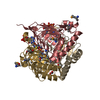
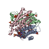
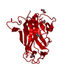
- PDB-3ci3: Structure of the PduO-type ATP:co(I)rrinoid adenosyltransferase f... -
+ Open data

- Basic information
| Entry | Database: PDB / ID: 3ci3 | ||||||
|---|---|---|---|---|---|---|---|
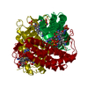
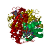
| Title | Structure of the PduO-type ATP:co(I)rrinoid adenosyltransferase from Lactobacillus reuteri complexed with partial adenosylcobalamin and PPPi | ||||||
 Components Components | Cobalamin adenosyltransferase PduO-like protein | ||||||
 Keywords Keywords |  TRANSFERASE / adenosyltransferase variant / Adenosylcobalamin binding / ATP binding TRANSFERASE / adenosyltransferase variant / Adenosylcobalamin binding / ATP binding | ||||||
| Function / homology |  Function and homology information Function and homology informationcorrinoid adenosyltransferase / corrinoid adenosyltransferase activity / cobalamin biosynthetic process / ATP binding / metal ion bindingSimilarity search - Function | ||||||
| Biological species |  Lactobacillus reuteri (bacteria) Lactobacillus reuteri (bacteria) | ||||||
| Method | X-RAY DIFFRACTION / SYNCHROTRON / Resolution: 1.11 Å | ||||||
 Authors Authors | Maurice, M.St. / Mera, P.E. / Escalante-Semerena, J.C. / Rayment, I. | ||||||
 Citation Citation | Journal: Biochemistry / Year: 2008 Title: Structural characterization of a human-type corrinoid adenosyltransferase confirms that coenzyme B12 is synthesized through a four-coordinate intermediate. Authors: St Maurice, M. / Mera, P. / Park, K. / Brunold, T.C. / Escalante-Semerena, J.C. / Rayment, I. | ||||||
| History |
|
- Structure visualization
| Structure viewer | Molecule: MolmilJmol/JSmol |
|---|
- Downloads & links
-Download
| PDBx/mmCIF format | 3ci3.cif.gz | 64 KB | Display | PDBx/mmCIF format |
|---|---|---|---|---|
| PDB format | pdb3ci3.ent.gz | 44.2 KB | Display | PDB format |
| PDBx/mmJSON format | 3ci3.json.gz | Tree view | PDBx/mmJSON format | |
| Others |  Other downloads Other downloads |
-Validation report
| Arichive directory | https://data.pdbj.org/pub/pdb/validation_reports/ci/3ci3ftp://data.pdbj.org/pub/pdb/validation_reports/ci/3ci3 | HTTPS FTP |
|---|
-Related structure data


-Links
 PDBj
PDBj

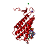
- Assembly
| Deposited unit | 
| |||||||||
|---|---|---|---|---|---|---|---|---|---|---|
| 1 | 
| |||||||||
| Unit cell |
| |||||||||
| Components on special symmetry positions |
|
-Components
-Protein , 1 types, 1 molecules A
| #1: Protein | Mass: 22382.316 Da / Num. of mol.: 1 Source method: isolated from a genetically manipulated source Source: (gene. exp.) Lactobacillus reuteri (bacteria) / Strain: CRL1098 / Gene: cobA / Plasmid: pET28B / Production host: Escherichia coli (E. coli) / Strain (production host): BL21(DE3) / References: UniProt: Q50EJ2, corrinoid adenosyltransferase |
|---|
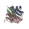
-Non-polymers , 6 types, 177 molecules 


| #2: Chemical | ChemComp-NA /  Mass: 22.990 Da / Num. of mol.: 1 / Source method: obtained synthetically / Formula: Na Mass: 22.990 Da / Num. of mol.: 1 / Source method: obtained synthetically / Formula: Na |
|---|---|
| #3: Chemical | ChemComp-MG /  Mass: 24.305 Da / Num. of mol.: 1 / Source method: obtained synthetically / Formula: Mg Mass: 24.305 Da / Num. of mol.: 1 / Source method: obtained synthetically / Formula: Mg |
| #4: Chemical | ChemComp-3PO / Polyphosphate Mass: 257.955 Da / Num. of mol.: 1 / Source method: obtained synthetically / Formula: H5O10P3 Mass: 257.955 Da / Num. of mol.: 1 / Source method: obtained synthetically / Formula: H5O10P3 |
| #5: Chemical | ChemComp-5AD / Deoxyadenosine Mass: 251.242 Da / Num. of mol.: 1 / Source method: obtained synthetically / Formula: C10H13N5O3 Mass: 251.242 Da / Num. of mol.: 1 / Source method: obtained synthetically / Formula: C10H13N5O3 |
| #6: Chemical | ChemComp-B12 / Vitamin B12 Mass: 1330.356 Da / Num. of mol.: 1 / Source method: obtained synthetically / Formula: C62H89CoN13O14P Mass: 1330.356 Da / Num. of mol.: 1 / Source method: obtained synthetically / Formula: C62H89CoN13O14P |
| #7: Water | ChemComp-HOH / WaterMass: 18.015 Da / Num. of mol.: 172 / Source method: isolated from a natural source / Formula: H2O |
-Experimental details
-Experiment
| Experiment | Method: X-RAY DIFFRACTION / Number of used crystals: 1 |
|---|
- Sample preparation
| Crystal | Density Matthews: 2.2 Å3/Da / Density % sol: 44.17 % |
|---|---|
| Crystal grow | Temperature: 300 K / Method: vapor diffusion / pH: 6 Details: ANOXIC, 10% PEG 8000, 0.1 M MES, 33 ug/mL FMN reductase, 20 mM NADH, 2 mM FMN, 2 mM hydroxycobalamin, 3 mM MgCl2, 3 mM ATP. Grown 65 days, VAPOR DIFFUSION, temperature 300K |
-Data collection
| Diffraction | Mean temperature: 100 K |
|---|---|
| Diffraction source | Source: SYNCHROTRON / Site: APS  / Beamline: 19-ID / Wavelength: 1.541 Å / Beamline: 19-ID / Wavelength: 1.541 Å |
| Detector | Type: ADSC QUANTUM 315 / Detector: CCD / Date: Dec 6, 2006 |
| Radiation | Protocol: SINGLE WAVELENGTH / Monochromatic (M) / Laue (L): M / Scattering type: x-ray |
| Radiation wavelength | Wavelength: 1.541 Å / Relative weight: 1 |
| Reflection | Resolution: 1.11→30 Å / Num. obs: 74669 / % possible obs: 98.9 % / Redundancy: 4.9 % / Biso Wilson estimate: 13.9 Å2 / Rmerge(I) obs: 0.39 / Net I/σ(I): 35.3 |
| Reflection shell | Resolution: 1.11→1.15 Å / Redundancy: 2.3 % / Rmerge(I) obs: 0.96 / Mean I/σ(I) obs: 9.8 / % possible all: 89 |
- Processing
| Software |
| ||||||||||||||||||||||||||||||||||||||||||||||||||||||||||||||||||||||||||||||||||||||||||
|---|---|---|---|---|---|---|---|---|---|---|---|---|---|---|---|---|---|---|---|---|---|---|---|---|---|---|---|---|---|---|---|---|---|---|---|---|---|---|---|---|---|---|---|---|---|---|---|---|---|---|---|---|---|---|---|---|---|---|---|---|---|---|---|---|---|---|---|---|---|---|---|---|---|---|---|---|---|---|---|---|---|---|---|---|---|---|---|---|---|---|---|
| Refinement | Resolution: 1.11→30 Å / Cor.coef. Fo:Fc: 0.974 / Cor.coef. Fo:Fc free: 0.965 / SU B: 0.387 / SU ML: 0.02 / Cross valid method: THROUGHOUT / σ(F): 0 / ESU R: 0.031 / ESU R Free: 0.033 / Stereochemistry target values: MAXIMUM LIKELIHOOD / Details: HYDROGENS HAVE BEEN ADDED IN THE RIDING POSITIONS
| ||||||||||||||||||||||||||||||||||||||||||||||||||||||||||||||||||||||||||||||||||||||||||
| Solvent computation | Ion probe radii: 0.8 Å / Shrinkage radii: 0.8 Å / VDW probe radii: 1.2 Å / Solvent model: MASK | ||||||||||||||||||||||||||||||||||||||||||||||||||||||||||||||||||||||||||||||||||||||||||
| Displacement parameters | Biso mean: 12.935 Å2
| ||||||||||||||||||||||||||||||||||||||||||||||||||||||||||||||||||||||||||||||||||||||||||
| Refine analyze | Luzzati coordinate error obs: 0.106 Å | ||||||||||||||||||||||||||||||||||||||||||||||||||||||||||||||||||||||||||||||||||||||||||
| Refinement step | Cycle: LAST / Resolution: 1.11→30 Å
| ||||||||||||||||||||||||||||||||||||||||||||||||||||||||||||||||||||||||||||||||||||||||||
| Refine LS restraints |
| ||||||||||||||||||||||||||||||||||||||||||||||||||||||||||||||||||||||||||||||||||||||||||
| LS refinement shell | Resolution: 1.11→1.143 Å / Total num. of bins used: 20
|
