 Movie
Movie Controller

[English] 日本語
 Yorodumi
- PDB-3c9e: Crystal structure of the cathepsin K : chondroitin sulfate complex. -
+ Open data

- Basic information
| Entry | Database: PDB / ID: 3c9e | |||||||||
|---|---|---|---|---|---|---|---|---|---|---|
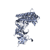
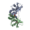
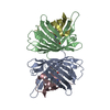
| Title | Crystal structure of the cathepsin K : chondroitin sulfate complex. | |||||||||
 Components Components | Cathepsin K | |||||||||
 Keywords Keywords | HYDROLASE / n:1 cathepsin K : chondroitin sulfate complex / "beads-on-a-string" organization / Lysosome / Protease / Thiol protease | |||||||||
| Function / homology |  Function and homology informationcathepsin K / mononuclear cell differentiation / intramembranous ossification / negative regulation of cartilage development / cellular response to zinc ion starvation / RUNX1 regulates transcription of genes involved in differentiation of keratinocytes / thyroid hormone generation / endolysosome lumen / Trafficking and processing of endosomal TLR / proteoglycan binding ...cathepsin K / mononuclear cell differentiation / intramembranous ossification / negative regulation of cartilage development / cellular response to zinc ion starvation / RUNX1 regulates transcription of genes involved in differentiation of keratinocytes / thyroid hormone generation / endolysosome lumen / Trafficking and processing of endosomal TLR / proteoglycan binding / Activation of Matrix Metalloproteinases / cysteine-type endopeptidase activator activity involved in apoptotic process / mitophagy / fibronectin binding / Collagen degradation / collagen catabolic process / extracellular matrix disassembly / bone resorption / cysteine-type peptidase activity / cellular response to transforming growth factor beta stimulus / collagen binding / MHC class II antigen presentation / Degradation of the extracellular matrix / lysosomal lumen / proteolysis involved in protein catabolic process / positive regulation of apoptotic signaling pathway / response to insulin / response to organic cyclic compound / cellular response to tumor necrosis factor / response to ethanol / lysosome / immune response / apical plasma membrane / external side of plasma membrane / cysteine-type endopeptidase activity / serine-type endopeptidase activity / intracellular membrane-bounded organelle / proteolysis / extracellular space / extracellular region / nucleoplasm Function and homology informationcathepsin K / mononuclear cell differentiation / intramembranous ossification / negative regulation of cartilage development / cellular response to zinc ion starvation / RUNX1 regulates transcription of genes involved in differentiation of keratinocytes / thyroid hormone generation / endolysosome lumen / Trafficking and processing of endosomal TLR / proteoglycan binding ...cathepsin K / mononuclear cell differentiation / intramembranous ossification / negative regulation of cartilage development / cellular response to zinc ion starvation / RUNX1 regulates transcription of genes involved in differentiation of keratinocytes / thyroid hormone generation / endolysosome lumen / Trafficking and processing of endosomal TLR / proteoglycan binding / Activation of Matrix Metalloproteinases / cysteine-type endopeptidase activator activity involved in apoptotic process / mitophagy / fibronectin binding / Collagen degradation / collagen catabolic process / extracellular matrix disassembly / bone resorption / cysteine-type peptidase activity / cellular response to transforming growth factor beta stimulus / collagen binding / MHC class II antigen presentation / Degradation of the extracellular matrix / lysosomal lumen / proteolysis involved in protein catabolic process / positive regulation of apoptotic signaling pathway / response to insulin / response to organic cyclic compound / cellular response to tumor necrosis factor / response to ethanol / lysosome / immune response / apical plasma membrane / external side of plasma membrane / cysteine-type endopeptidase activity / serine-type endopeptidase activity / intracellular membrane-bounded organelle / proteolysis / extracellular space / extracellular region / nucleoplasmSimilarity search - Function | |||||||||
| Biological species |  Homo sapiens (human) Homo sapiens (human) | |||||||||
| Method | X-RAY DIFFRACTION / SYNCHROTRON / Resolution: 1.8 Å | |||||||||
 Authors Authors | Kienetz, M. / Cherney, M.M. / James, M.N.G. / Bromme, D. | |||||||||
 Citation Citation | Journal: J.Mol.Biol. / Year: 2008 Title: The crystal and molecular structures of a cathepsin K:chondroitin sulfate complex. Authors: Li, Z. / Kienetz, M. / Cherney, M.M. / James, M.N. / Bromme, D. | |||||||||
| History |
|
- Structure visualization
| Structure viewer | Molecule: MolmilJmol/JSmol |
|---|
- Downloads & links
-Download
| PDBx/mmCIF format | 3c9e.cif.gz | 61.2 KB | Display | PDBx/mmCIF format |
|---|---|---|---|---|
| PDB format | pdb3c9e.ent.gz | 47.2 KB | Display | PDB format |
| PDBx/mmJSON format | 3c9e.json.gz | Tree view | PDBx/mmJSON format | |
| Others |  Other downloads Other downloads |
-Validation report
| Arichive directory | https://data.pdbj.org/pub/pdb/validation_reports/c9/3c9eftp://data.pdbj.org/pub/pdb/validation_reports/c9/3c9e | HTTPS FTP |
|---|
-Related structure data
| Similar structure data |
|---|


-Links
 PDBj
PDBj


- Assembly
| Deposited unit | 
| ||||||||||||
|---|---|---|---|---|---|---|---|---|---|---|---|---|---|
| 1 | 
| ||||||||||||
| Unit cell |
| ||||||||||||
| Components on special symmetry positions |
| ||||||||||||
| Details | Author states that biological assembly contains one chondroitin sulfate molecule and N cathepsin K molecules. Depending on their ratio in solution and the Chondroitin sulfate moleculat weight N can vary from 1 to 20 molecules. |
-Components
| #1: Protein | / Cathepsin O / Cathepsin X / Cathepsin O2 Mass: 23523.480 Da / Num. of mol.: 1 Source method: isolated from a genetically manipulated source Source: (gene. exp.) Homo sapiens (human) / Gene: CTSK, CTSO, CTSO2 / Production host:  Pichia pastoris (fungus) / References: UniProt: P43235, cathepsin K Pichia pastoris (fungus) / References: UniProt: P43235, cathepsin K |
|---|---|
| #2: Polysaccharide | 2-acetamido-2-deoxy-4-O-sulfo-beta-D-galactopyranose-(1-4)-beta-D-glucopyranuronic acid-(1-3)-2- ...2-acetamido-2-deoxy-4-O-sulfo-beta-D-galactopyranose-(1-4)-beta-D-glucopyranuronic acid-(1-3)-2-acetamido-2-deoxy-4-O-sulfo-beta-D-galactopyranose-(1-4)-beta-D-glucopyranuronic acid-(1-3)-2-acetamido-2-deoxy-4-O-sulfo-beta-D-galactopyranose-(1-4)-beta-D-glucopyranuronic acid / Mass: 1396.154 Da / Num. of mol.: 1 Source method: isolated from a genetically manipulated source |
| #3: Chemical | ChemComp-CA /   Mass: 40.078 Da / Num. of mol.: 1 / Source method: obtained synthetically / Formula: Ca Mass: 40.078 Da / Num. of mol.: 1 / Source method: obtained synthetically / Formula: Ca |
| #4: Chemical | ChemComp-E64 /   Mass: 360.429 Da / Num. of mol.: 1 / Source method: obtained synthetically / Formula: C15H30N5O5 Mass: 360.429 Da / Num. of mol.: 1 / Source method: obtained synthetically / Formula: C15H30N5O5 |
| #5: Water | ChemComp-HOH / Water Mass: 18.015 Da / Num. of mol.: 310 / Source method: isolated from a natural source / Formula: H2O Mass: 18.015 Da / Num. of mol.: 310 / Source method: isolated from a natural source / Formula: H2O |
-Experimental details
-Experiment
| Experiment | Method: X-RAY DIFFRACTION / Number of used crystals: 1 |
|---|
- Sample preparation
| Crystal | Density Matthews: 2.8 Å3/Da / Density % sol: 56.08 % |
|---|---|
| Crystal grow | Temperature: 295 K / Method: vapor diffusion, hanging drop / pH: 4.5 Details: Cathepsin K:chondroitin sulfate complex was made at 1:1 ratio. Precipitant contained 30% MPD, 0.1M sodium acetate buffer, 20mM calcium chloride, pH 4.5, VAPOR DIFFUSION, HANGING DROP, ...Details: Cathepsin K:chondroitin sulfate complex was made at 1:1 ratio. Precipitant contained 30% MPD, 0.1M sodium acetate buffer, 20mM calcium chloride, pH 4.5, VAPOR DIFFUSION, HANGING DROP, temperature 295K, VAPOR DIFFUSION, HANGING DROP |
-Data collection
| Diffraction source | Source: SYNCHROTRON / Site: ALS  / Beamline: 8.3.1 / Wavelength: 1.05 Å / Beamline: 8.3.1 / Wavelength: 1.05 Å |
|---|---|
| Detector | Type: ADSC QUANTUM 210 / Detector: CCD / Date: Mar 2, 2002 |
| Radiation | Protocol: SINGLE WAVELENGTH / Monochromatic (M) / Laue (L): M / Scattering type: x-ray |
| Radiation wavelength | Wavelength: 1.05 Å / Relative weight: 1 |
| Reflection | Resolution: 1.8→40 Å / Num. all: 25031 / Num. obs: 25031 / % possible obs: 99.8 % / Observed criterion σ(F): 0 / Observed criterion σ(I): 0 / Redundancy: 4 % / Rmerge(I) obs: 0.142 / Net I/σ(I): 7.3 |
| Reflection shell | Resolution: 1.8→1.9 Å / Redundancy: 4.1 % / Rmerge(I) obs: 0.742 / Mean I/σ(I) obs: 1.9 / % possible all: 99.7 |
- Processing
| Software |
| ||||||||||||||||||||||||||||||||||||||||||||||||||||||||||||||||||||||||||||||||
|---|---|---|---|---|---|---|---|---|---|---|---|---|---|---|---|---|---|---|---|---|---|---|---|---|---|---|---|---|---|---|---|---|---|---|---|---|---|---|---|---|---|---|---|---|---|---|---|---|---|---|---|---|---|---|---|---|---|---|---|---|---|---|---|---|---|---|---|---|---|---|---|---|---|---|---|---|---|---|---|---|---|
| Refinement | Resolution: 1.8→40 Å / σ(F): 0
| ||||||||||||||||||||||||||||||||||||||||||||||||||||||||||||||||||||||||||||||||
| Solvent computation | Bsol: 61.68 Å2 | ||||||||||||||||||||||||||||||||||||||||||||||||||||||||||||||||||||||||||||||||
| Displacement parameters | Biso mean: 17.098 Å2
| ||||||||||||||||||||||||||||||||||||||||||||||||||||||||||||||||||||||||||||||||
| Refinement step | Cycle: LAST / Resolution: 1.8→40 Å
| ||||||||||||||||||||||||||||||||||||||||||||||||||||||||||||||||||||||||||||||||
| Refine LS restraints |
| ||||||||||||||||||||||||||||||||||||||||||||||||||||||||||||||||||||||||||||||||
| Xplor file |
|
