- PDB-3c8e: Crystal Structure Analysis of yghU from E. Coli -
+
Open data
ID or keywords:
Loading...
-
Basic information
Entry
Database: PDB / ID: 3c8e
Title
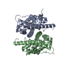
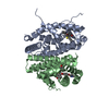
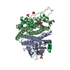
Crystal Structure Analysis of yghU from E. Coli
Components
yghU, glutathione S-transferase homologue
Keywords
TRANSFERASE / glutathione transferase homologue / YghU / E. coli
Function / homology
Function and homology information
Oxidoreductases; Acting on a sulfur group of donors; With a disulfide as acceptor / disulfide oxidoreductase activity / Oxidoreductases; Acting on a peroxide as acceptor; Peroxidases / peroxidase activity Similarity search - Function
Redundancy: 6.6 % / Av σ(I) over netI: 9.4 / Number: 295858 / Rmerge(I) obs: 0.097 / Χ2: 1 / D res high: 1.89 Å / D res low: 50 Å / Num. obs: 44647 / % possible obs: 96.7
Diffraction reflection shell
Highest resolution (Å)
Lowest resolution (Å)
% possible obs (%)
ID
Rmerge(I) obs
Chi squared
Redundancy
4.07
50
99.8
1
0.061
0.994
7.7
3.23
4.07
100
1
0.077
0.925
7.6
2.82
3.23
99.9
1
0.088
0.924
7.1
2.57
2.82
99.6
1
0.112
0.965
6.8
2.38
2.57
99.5
1
0.139
1.032
6.8
2.24
2.38
98.8
1
0.176
1.058
6.7
2.13
2.24
98
1
0.22
1.053
6.6
2.04
2.13
96.8
1
0.271
1.099
6.3
1.96
2.04
94.8
1
0.332
0.962
5.7
1.89
1.96
79.5
1
0.392
0.929
4.2
Reflection
Resolution: 1.41→50 Å / Num. obs: 95609 / % possible obs: 87.7 % / Redundancy: 8.7 % / Rmerge(I) obs: 0.109 / Χ2: 1.039 / Net I/σ(I): 7.1
Reflection shell
Resolution (Å)
Redundancy (%)
Rmerge(I) obs
Num. unique all
Χ2
% possible all
1.41-1.46
3
0.653
6087
0.724
56.5
1.46-1.52
3.9
0.567
7908
0.654
73.4
1.52-1.59
4.7
0.517
8596
0.748
79.7
1.59-1.67
5.6
0.488
9185
0.741
85
1.67-1.78
7.6
0.528
9837
0.787
91
1.78-1.91
9.8
0.482
10310
0.802
95.1
1.91-2.11
10.8
0.299
10570
0.857
97.3
2.11-2.41
11.2
0.175
10798
0.961
98.7
2.41-3.04
11.6
0.108
10957
1.125
99.5
3.04-50
13.1
0.055
11361
1.693
99.6
-
Phasing
Phasing
Method: MAD
-
Processing
Software
Name
Version
Classification
NB
DENZO
datareduction
SCALEPACK
datascaling
SHELX
phasing
REFMAC
refinement
PDB_EXTRACT
3.004
dataextraction
HKL-2000
datareduction
HKL-2000
datascaling
SHELXD
phasing
Refinement
Method to determine structure: MAD / Resolution: 1.5→20 Å / Cor.coef. Fo:Fc: 0.967 / Cor.coef. Fo:Fc free: 0.952 / SU B: 1.274 / SU ML: 0.048 / Cross valid method: THROUGHOUT / σ(F): 0 / ESU R: 0.08 / ESU R Free: 0.082 / Stereochemistry target values: MAXIMUM LIKELIHOOD
Rfactor
Num. reflection
% reflection
Selection details
Rfree
0.197
8037
10 %
RANDOM
Rwork
0.162
-
-
-
obs
0.165
80150
100 %
-
Solvent computation
Ion probe radii: 0.8 Å / Shrinkage radii: 0.8 Å / VDW probe radii: 1.2 Å / Solvent model: BABINET MODEL WITH MASK
Displacement parameters
Biso mean: 14.609 Å2
Baniso -1
Baniso -2
Baniso -3
1-
0.24 Å2
0 Å2
0 Å2
2-
-
-0.98 Å2
0 Å2
3-
-
-
0.73 Å2
Refinement step
Cycle: LAST / Resolution: 1.5→20 Å
Protein
Nucleic acid
Ligand
Solvent
Total
Num. atoms
4523
0
80
471
5074
Refine LS restraints
Refine-ID
Type
Dev ideal
Dev ideal target
Number
X-RAY DIFFRACTION
r_bond_refined_d
0.019
0.022
4882
X-RAY DIFFRACTION
r_angle_refined_deg
1.834
1.953
6662
X-RAY DIFFRACTION
r_dihedral_angle_1_deg
5.864
5
611
X-RAY DIFFRACTION
r_dihedral_angle_2_deg
32.997
23.852
244
X-RAY DIFFRACTION
r_dihedral_angle_3_deg
13.133
15
776
X-RAY DIFFRACTION
r_dihedral_angle_4_deg
14.292
15
31
X-RAY DIFFRACTION
r_chiral_restr
0.247
0.2
691
X-RAY DIFFRACTION
r_gen_planes_refined
0.01
0.02
3873
X-RAY DIFFRACTION
r_nbd_refined
0.22
0.2
2321
X-RAY DIFFRACTION
r_nbtor_refined
0.315
0.2
3360
X-RAY DIFFRACTION
r_xyhbond_nbd_refined
0.128
0.2
450
X-RAY DIFFRACTION
r_symmetry_vdw_refined
0.198
0.2
47
X-RAY DIFFRACTION
r_symmetry_hbond_refined
0.213
0.2
22
X-RAY DIFFRACTION
r_mcbond_it
1.191
1.5
2957
X-RAY DIFFRACTION
r_mcangle_it
1.677
2
4651
X-RAY DIFFRACTION
r_scbond_it
2.612
3
2221
X-RAY DIFFRACTION
r_scangle_it
3.826
4.5
1988
LS refinement shell
Resolution: 1.5→1.581 Å / Total num. of bins used: 10
Rfactor
Num. reflection
% reflection
Rfree
0.272
882
-
Rwork
0.215
8027
-
all
-
8909
-
obs
-
-
100 %
+
About Yorodumi
-
News
-
Feb 9, 2022. New format data for meta-information of EMDB entries
New format data for meta-information of EMDB entries
Version 3 of the EMDB header file is now the official format.
The previous official version 1.9 will be removed from the archive.
In the structure databanks used in Yorodumi, some data are registered as the other names, "COVID-19 virus" and "2019-nCoV". Here are the details of the virus and the list of structure data.
Jan 31, 2019. EMDB accession codes are about to change! (news from PDBe EMDB page)
EMDB accession codes are about to change! (news from PDBe EMDB page)
The allocation of 4 digits for EMDB accession codes will soon come to an end. Whilst these codes will remain in use, new EMDB accession codes will include an additional digit and will expand incrementally as the available range of codes is exhausted. The current 4-digit format prefixed with “EMD-” (i.e. EMD-XXXX) will advance to a 5-digit format (i.e. EMD-XXXXX), and so on. It is currently estimated that the 4-digit codes will be depleted around Spring 2019, at which point the 5-digit format will come into force.
The EM Navigator/Yorodumi systems omit the EMD- prefix.
Related info.:Q: What is EMD? / ID/Accession-code notation in Yorodumi/EM Navigator
Yorodumi is a browser for structure data from EMDB, PDB, SASBDB, etc.
This page is also the successor to EM Navigator detail page, and also detail information page/front-end page for Omokage search.
The word "yorodu" (or yorozu) is an old Japanese word meaning "ten thousand". "mi" (miru) is to see.
Related info.:EMDB / PDB / SASBDB / Comparison of 3 databanks / Yorodumi Search / Aug 31, 2016. New EM Navigator & Yorodumi / Yorodumi Papers / Jmol/JSmol / Function and homology information / Changes in new EM Navigator and Yorodumi
 Movie
Movie Controller


 Open data
 Basic information
 Components
 Keywords
 TRANSFERASE / glutathione transferase homologue / YghU /
  Function and homology information
 Authors
 Citation
 Structure visualization
 Downloads & links
 Other downloads
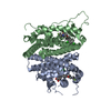


 PDBj
PDBj

 Assembly

 Mass: 307.323 Da / Num. of mol.: 4 / Source method: obtained synthetically / Formula: C10H17N3O6S
 Mass: 18.015 Da / Num. of mol.: 471 / Source method: isolated from a natural source / Formula: H2O
 Sample preparation
 / Beamline: 22-BM / Wavelength: 1.00, 0.9795, 0.9790, 0.9500
 Processing