 Movie
Movie Controller

[English] 日本語
 Yorodumi
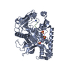
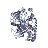
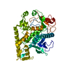
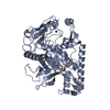
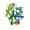
- PDB-3c8b: Crystal structure of the catalytic domain of botulinum neurotoxin... -
+ Open data

- Basic information
| Entry | Database: PDB / ID: 3c8b | ||||||
|---|---|---|---|---|---|---|---|
| Title | Crystal structure of the catalytic domain of botulinum neurotoxin serotype A with inhibitory peptide RRGI | ||||||
 Components Components |
| ||||||
 Keywords Keywords | HYDROLASE/HYDROLASE INHIBITOR / BOTULINUM NEUROTOXIN TYPE A /  CATALYTIC DOMAIN / ENDOPEPTIDASE / BIO-WARFARE AGENT / METALLOPROTEASE / PROTEASE / SECRETED / HYDROLASE-HYDROLASE INHIBITOR complex CATALYTIC DOMAIN / ENDOPEPTIDASE / BIO-WARFARE AGENT / METALLOPROTEASE / PROTEASE / SECRETED / HYDROLASE-HYDROLASE INHIBITOR complex | ||||||
| Function / homology |  Function and homology information Function and homology informationhost cell junction / negative regulation of neurotransmitter secretion / bontoxilysin / host cell presynaptic membrane / host cell cytoplasmic vesicle / host cell cytosol / protein transmembrane transporter activity / metalloendopeptidase activity / toxin activity / membrane => GO:0016020 ...host cell junction / negative regulation of neurotransmitter secretion / bontoxilysin / host cell presynaptic membrane / host cell cytoplasmic vesicle / host cell cytosol / protein transmembrane transporter activity / metalloendopeptidase activity / toxin activity / membrane => GO:0016020 / host cell plasma membrane / proteolysis / zinc ion binding / extracellular region / membraneSimilarity search - Function | ||||||
| Biological species |   Clostridium botulinum (bacteria) Clostridium botulinum (bacteria) | ||||||
| Method | X-RAY DIFFRACTION / SYNCHROTRON / MOLECULAR REPLACEMENT / Resolution: 1.47 Å | ||||||
 Authors Authors | Kumaran, D. / Swaminathan, S. | ||||||
 Citation Citation | Journal: J.Biol.Chem. / Year: 2008 Title: Structure- and Substrate-based Inhibitor Design for Clostridium botulinum Neurotoxin Serotype A Authors: Kumaran, D. / Rawat, R. / Ludivico, M.L. / Ahmed, S.A. / Swaminathan, S. | ||||||
| History |
|
- Structure visualization
| Structure viewer | Molecule: MolmilJmol/JSmol |
|---|
- Downloads & links
-Download
| PDBx/mmCIF format | 3c8b.cif.gz | 107.6 KB | Display | PDBx/mmCIF format |
|---|---|---|---|---|
| PDB format | pdb3c8b.ent.gz | 81.2 KB | Display | PDB format |
| PDBx/mmJSON format | 3c8b.json.gz | Tree view | PDBx/mmJSON format | |
| Others |  Other downloads Other downloads |
-Validation report
| Arichive directory | https://data.pdbj.org/pub/pdb/validation_reports/c8/3c8bftp://data.pdbj.org/pub/pdb/validation_reports/c8/3c8b | HTTPS FTP |
|---|
-Related structure data
| Related structure data | 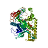 3bwiSC  3c88C 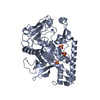 3c89C  3c8aC S: Starting model for refinement C: citing same article ( |
|---|---|
| Similar structure data |

-Links
 PDBj
PDBj- Assembly
| Deposited unit | 
| ||||||||
|---|---|---|---|---|---|---|---|---|---|
| 1 |
| ||||||||
| Unit cell |
|
-Components
| #1: Protein | Mass: 49657.996 Da / Num. of mol.: 1 / Fragment: RESIDUES 1-424 Source method: isolated from a genetically manipulated source Source: (gene. exp.) Clostridium botulinum (bacteria) / Strain: HALL / Gene: botA / Plasmid: PET28B / Production host: Escherichia coli (E. coli) / Strain (production host): BL21-DE3 RILReferences: UniProt: A5HZZ9, UniProt: P0DPI1*PLUS, bontoxilysin | ||
|---|---|---|---|
| #2: Protein/peptide | Mass: 500.620 Da / Num. of mol.: 1 / Source method: obtained synthetically | ||
| #3: Chemical | ChemComp-ZN /   Mass: 65.409 Da / Num. of mol.: 1 / Source method: obtained synthetically / Formula: Zn Mass: 65.409 Da / Num. of mol.: 1 / Source method: obtained synthetically / Formula: Zn | ||
| #4: Chemical | Sulfate  Mass: 96.063 Da / Num. of mol.: 3 / Source method: obtained synthetically / Formula: SO4 Mass: 96.063 Da / Num. of mol.: 3 / Source method: obtained synthetically / Formula: SO4#5: Water | ChemComp-HOH / | Water Mass: 18.015 Da / Num. of mol.: 372 / Source method: isolated from a natural source / Formula: H2O Mass: 18.015 Da / Num. of mol.: 372 / Source method: isolated from a natural source / Formula: H2O |
-Experimental details
-Experiment
| Experiment | Method: X-RAY DIFFRACTION / Number of used crystals: 1 |
|---|
- Sample preparation
| Crystal | Density Matthews: 2.19 Å3/Da / Density % sol: 43.77 % |
|---|---|
| Crystal grow | Temperature: 293 K / Method: vapor diffusion, sitting drop / pH: 6.9 Details: 17% PEG 3350, 0.25M AMMONIUM SULFATE, 0.1M BIS-TRIS, pH 6.90, VAPOR DIFFUSION, SITTING DROP, temperature 293K |
-Data collection
| Diffraction | Mean temperature: 100 K |
|---|---|
| Diffraction source | Source: SYNCHROTRON / Site: NSLS  / Beamline: X29A / Wavelength: 0.979 / Wavelength: 0.979 Å / Beamline: X29A / Wavelength: 0.979 / Wavelength: 0.979 Å |
| Detector | Type: ADSC QUANTUM 315 / Detector: CCD / Date: Oct 22, 2007 / Details: MIRRORS |
| Radiation | Monochromator: SI 111 / Protocol: SINGLE WAVELENGTH / Monochromatic (M) / Laue (L): M / Scattering type: x-ray |
| Radiation wavelength | Wavelength: 0.979 Å / Relative weight: 1 |
| Reflection | Resolution: 1.47→50 Å / Num. all: 63031 / Num. obs: 63031 / % possible obs: 87 % / Observed criterion σ(F): 0 / Observed criterion σ(I): 0 / Redundancy: 15.1 % / Biso Wilson estimate: 14.5 Å2 / Rmerge(I) obs: 0.066 / Net I/σ(I): 20 |
| Reflection shell | Resolution: 1.47→1.51 Å / Redundancy: 5.2 % / Rmerge(I) obs: 0.194 / Mean I/σ(I) obs: 3 / Num. unique all: 3135 / % possible all: 52.2 |
- Processing
| Software |
| ||||||||||||||||||||||||||||||||||||||||||||||||||||||||||||
|---|---|---|---|---|---|---|---|---|---|---|---|---|---|---|---|---|---|---|---|---|---|---|---|---|---|---|---|---|---|---|---|---|---|---|---|---|---|---|---|---|---|---|---|---|---|---|---|---|---|---|---|---|---|---|---|---|---|---|---|---|---|
| Refinement | Method to determine structure: MOLECULAR REPLACEMENT Starting model: 3BWI Resolution: 1.47→33.2 Å / Rfactor Rfree error: 0.004 / Data cutoff high absF: 79982.79 / Data cutoff low absF: 0 / Isotropic thermal model: RESTRAINED / Cross valid method: THROUGHOUT / σ(F): 0 / Stereochemistry target values: Engh & Huber
| ||||||||||||||||||||||||||||||||||||||||||||||||||||||||||||
| Solvent computation | Solvent model: FLAT MODEL / Bsol: 41.3764 Å2 / ksol: 0.363297 e/Å3 | ||||||||||||||||||||||||||||||||||||||||||||||||||||||||||||
| Displacement parameters | Biso mean: 23.1 Å2
| ||||||||||||||||||||||||||||||||||||||||||||||||||||||||||||
| Refine analyze |
| ||||||||||||||||||||||||||||||||||||||||||||||||||||||||||||
| Refinement step | Cycle: LAST / Resolution: 1.47→33.2 Å
| ||||||||||||||||||||||||||||||||||||||||||||||||||||||||||||
| Refine LS restraints |
| ||||||||||||||||||||||||||||||||||||||||||||||||||||||||||||
| LS refinement shell | Resolution: 1.47→1.56 Å / Rfactor Rfree error: 0.015 / Total num. of bins used: 6
|
