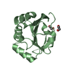
| Deposited unit | A: Thiol-disulfide oxidoreductase resA
B: Thiol-disulfide oxidoreductase resA
hetero molecules
| Theoretical mass | Number of molelcules |
|---|
| Total (without water) | 31,516 | 3 |
|---|
| Polymers | 31,420 | 2 |
|---|
| Non-polymers | 96 | 1 |
|---|
| Water | 1,099 | 61 |
|---|
|
|---|
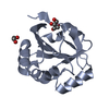
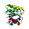
| 1 | A: Thiol-disulfide oxidoreductase resA
hetero molecules
| Theoretical mass | Number of molelcules |
|---|
| Total (without water) | 15,806 | 2 |
|---|
| Polymers | 15,710 | 1 |
|---|
| Non-polymers | 96 | 1 |
|---|
| Water | 18 | 1 |
|---|
| Type | Name | Symmetry operation | Number |
|---|
| identity operation | 1_555 | x,y,z | 1 |
|
|---|
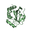
| 2 | B: Thiol-disulfide oxidoreductase resA
| Theoretical mass | Number of molelcules |
|---|
| Total (without water) | 15,710 | 1 |
|---|
| Polymers | 15,710 | 1 |
|---|
| Non-polymers | 0 | 0 |
|---|
| Water | 18 | 1 |
|---|
| Type | Name | Symmetry operation | Number |
|---|
| identity operation | 1_555 | x,y,z | 1 |
|
|---|
| Unit cell | | Length a, b, c (Å) | 47.219, 60.169, 110.393 |
|---|
| Angle α, β, γ (deg.) | 90.00, 90.00, 90.00 |
|---|
| Int Tables number | 19 |
|---|
| Space group name H-M | P212121 |
|---|
|
|---|
| Noncrystallographic symmetry (NCS) | NCS domain: | ID | Ens-ID | Details |
|---|
| 1 | 1 | A| 2 | 1 | B| 3 | 1 | A| 4 | 1 | B| 5 | 1 | A| 6 | 1 | B| 7 | 1 | A| 8 | 1 | B| 9 | 1 | A| 10 | 1 | B| 1 | 2 | B| 2 | 2 | A| 3 | 2 | B| 4 | 2 | A| 5 | 2 | B| 6 | 2 | A| 7 | 2 | B| 8 | 2 | A| 9 | 2 | B| 10 | 2 | A | | | | | | | | | | | | | | | | | | | |
NCS domain segments: Refine code: 5 | Dom-ID | Component-ID | Ens-ID | Beg auth comp-ID | Beg label comp-ID | End auth comp-ID | End label comp-ID | Auth asym-ID | Label asym-ID | Auth seq-ID | Label seq-ID |
|---|
| 1 | 1 | 1 | SERSERGLYGLYAA| 40 - 52 | 1 - 13 | | 2 | 1 | 1 | SERSERGLYGLYBB| 40 - 52 | 1 - 13 | | 3 | 2 | 1 | ARGARGASPASPAA| 54 - 59 | 15 - 20 | | 4 | 2 | 1 | ARGARGASPASPBB| 54 - 59 | 15 - 20 | | 5 | 3 | 1 | LYSLYSTHRTHRAA| 61 - 72 | 22 - 33 | | 6 | 3 | 1 | LYSLYSTHRTHRBB| 61 - 72 | 22 - 33 | | 7 | 4 | 1 | CYSCYSALAALAAA| 77 - 109 | 38 - 70 | | 8 | 4 | 1 | CYSCYSALAALABB| 77 - 109 | 38 - 70 | | 9 | 5 | 1 | HISHISGLYGLYAA| 111 - 175 | 72 - 136 | | 10 | 5 | 1 | HISHISGLYGLYBB| 111 - 175 | 72 - 136 | | 1 | 1 | 2 | SERSERGLYGLYBB| 40 - 52 | 1 - 13 | | 2 | 1 | 2 | SERSERGLYGLYAA| 40 - 52 | 1 - 13 | | 3 | 2 | 2 | ARGARGASPASPB| B | | | | | | | | | | | | | | | | | | | | | | | | | | | | | | | | | | | | | | | | | | | | | | | | | | | | | | | | | | | | | | | | | | | | | | | | | | | | | |
|
|---|
 Movie
Movie Controller


 Open data
 Basic information
 Components
 Keywords
 OXIDOREDUCTASE / Thioredoxin-like fold / Cytochrome c-type biogenesis /
  Function and homology information
 Authors
 Citation
 Structure visualization
 Downloads & links
 Other downloads
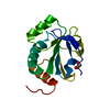


 PDBj
PDBj

 Assembly

