 Movie
Movie Controller

[English] 日本語
 Yorodumi
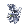
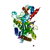
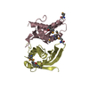
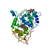
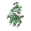
- PDB-3c4a: Crystal structure of vioD hydroxylase in complex with FAD from Ch... -
+ Open data

- Basic information
| Entry | Database: PDB / ID: 3c4a | ||||||
|---|---|---|---|---|---|---|---|
| Title | Crystal structure of vioD hydroxylase in complex with FAD from Chromobacterium violaceum. Northeast Structural Genomics Consortium Target CvR158 | ||||||
 Components Components | Probable tryptophan hydroxylase vioD | ||||||
 Keywords Keywords |  OXIDOREDUCTASE / alpha-beta protein / Structural Genomics / PSI-2 / Protein Structure Initiative / Northeast Structural Genomics Consortium / NESG / Antibiotic biosynthesis / FAD / Flavoprotein OXIDOREDUCTASE / alpha-beta protein / Structural Genomics / PSI-2 / Protein Structure Initiative / Northeast Structural Genomics Consortium / NESG / Antibiotic biosynthesis / FAD / Flavoprotein | ||||||
| Function / homology |  Function and homology information Function and homology informationprotodeoxyviolaceinate monooxygenase / antibiotic biosynthetic process / monooxygenase activitySimilarity search - Function | ||||||
| Biological species |  Chromobacterium violaceum ATCC 12472 (bacteria) Chromobacterium violaceum ATCC 12472 (bacteria) | ||||||
| Method | X-RAY DIFFRACTION / SYNCHROTRON / SAD / Resolution: 2.3 Å | ||||||
 Authors Authors | Forouhar, F. / Neely, H. / Seetharaman, J. / Janjua, H. / Xiao, R. / Maglaqui, M. / Wang, H. / Baran, M.C. / Acton, T.B. / Montelione, G.T. ...Forouhar, F. / Neely, H. / Seetharaman, J. / Janjua, H. / Xiao, R. / Maglaqui, M. / Wang, H. / Baran, M.C. / Acton, T.B. / Montelione, G.T. / Hunt, J.F. / Tong, L. / Northeast Structural Genomics Consortium (NESG) | ||||||
 Citation Citation | Journal: To be Published Title: Crystal structure of vioD hydroxylase in complex with FAD from Chromobacterium violaceum. Authors: Forouhar, F. / Neely, H. / Seetharaman, J. / Janjua, H. / Xiao, R. / Maglaqui, M. / Wang, H. / Baran, M.C. / Acton, T.B. / Montelione, G.T. / Hunt, J.F. / Tong, L. | ||||||
| History |
|
- Structure visualization
| Structure viewer | Molecule: MolmilJmol/JSmol |
|---|
- Downloads & links
-Download
| PDBx/mmCIF format | 3c4a.cif.gz | 84.1 KB | Display | PDBx/mmCIF format |
|---|---|---|---|---|
| PDB format | pdb3c4a.ent.gz | 67.7 KB | Display | PDB format |
| PDBx/mmJSON format | 3c4a.json.gz | Tree view | PDBx/mmJSON format | |
| Others |  Other downloads Other downloads |
-Validation report
| Arichive directory | https://data.pdbj.org/pub/pdb/validation_reports/c4/3c4aftp://data.pdbj.org/pub/pdb/validation_reports/c4/3c4a | HTTPS FTP |
|---|
-Related structure data
| Similar structure data | |
|---|---|
| Other databases |


-Links
 PDBj
PDBj- Assembly
| Deposited unit | 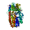
| ||||||||
|---|---|---|---|---|---|---|---|---|---|
| 1 |
| ||||||||
| Unit cell |
|
-Components
| #1: Protein | Mass: 43168.734 Da / Num. of mol.: 1 Source method: isolated from a genetically manipulated source Source: (gene. exp.) Chromobacterium violaceum ATCC 12472 (bacteria)Species: Chromobacterium violaceum / Strain: DSM 30191 / IFO 12614 / JCM 1249 / NCIB 9131 / Gene: vioD, CV_3271 / Plasmid: pET21 / Production host: Escherichia coli (E. coli) / Strain (production host): BL21(DE3)+Magic / References: UniProt: Q9S3U8, Oxidoreductases |
|---|---|
| #2: Chemical | ChemComp-FAD / Flavin adenine dinucleotide  Mass: 785.550 Da / Num. of mol.: 1 / Source method: obtained synthetically / Formula: C27H33N9O15P2 / Comment: FAD*YM Mass: 785.550 Da / Num. of mol.: 1 / Source method: obtained synthetically / Formula: C27H33N9O15P2 / Comment: FAD*YM |
| #3: Water | ChemComp-HOH / Water Mass: 18.015 Da / Num. of mol.: 86 / Source method: isolated from a natural source / Formula: H2O Mass: 18.015 Da / Num. of mol.: 86 / Source method: isolated from a natural source / Formula: H2O |
-Experimental details
-Experiment
| Experiment | Method: X-RAY DIFFRACTION / Number of used crystals: 1 |
|---|
- Sample preparation
| Crystal | Density Matthews: 2.05 Å3/Da / Density % sol: 39.98 % Description: The structure factor file contains Friedel pairs |
|---|---|
| Crystal grow | Temperature: 291 K / Method: vapor diffusion, sitting drop / pH: 6.5 Details: Protein solution: 10 mM Tris-HCl, 100 mM NaCl, 3 mM FAD, 5 mM DTT. Reservoir solution: 100 mM Bis-Tris pH 6.5, 25% PEG 3350, 200 mM MgCl2, VAPOR DIFFUSION, SITTING DROP, temperature 291K |
-Data collection
| Diffraction | Mean temperature: 100 K |
|---|---|
| Diffraction source | Source: SYNCHROTRON / Site: NSLS  / Beamline: X4C / Wavelength: 0.979 Å / Beamline: X4C / Wavelength: 0.979 Å |
| Detector | Type: MAR CCD 165 mm / Detector: CCD / Date: Jan 18, 2008 / Details: Mirrors |
| Radiation | Monochromator: Si 111 CHANNEL / Protocol: SINGLE WAVELENGTH / Monochromatic (M) / Laue (L): M / Scattering type: x-ray |
| Radiation wavelength | Wavelength: 0.979 Å / Relative weight: 1 |
| Reflection | Resolution: 2.3→30 Å / Num. all: 30479 / Num. obs: 30479 / % possible obs: 99.9 % / Observed criterion σ(F): 0 / Observed criterion σ(I): 0 / Redundancy: 7.5 % / Biso Wilson estimate: 18.4 Å2 / Rmerge(I) obs: 0.105 / Rsym value: 0.082 / Net I/σ(I): 20.58 |
| Reflection shell | Resolution: 2.3→2.38 Å / Redundancy: 7.4 % / Rmerge(I) obs: 0.473 / Mean I/σ(I) obs: 3.32 / Num. unique all: 3061 / Rsym value: 0.422 / % possible all: 100 |
- Processing
| Software |
| |||||||||||||||||||||||||
|---|---|---|---|---|---|---|---|---|---|---|---|---|---|---|---|---|---|---|---|---|---|---|---|---|---|---|
| Refinement | Method to determine structure: SAD / Resolution: 2.3→19.88 Å / Rfactor Rfree error: 0.005 / Data cutoff high absF: 345893.74 / Data cutoff low absF: 0 / Isotropic thermal model: OVERALL / Cross valid method: THROUGHOUT / σ(F): 2 / Stereochemistry target values: Engh & Huber / Details: The Friedel pairs were used in phasing
| |||||||||||||||||||||||||
| Solvent computation | Solvent model: FLAT MODEL / Bsol: 28.3601 Å2 / ksol: 0.35 e/Å3 | |||||||||||||||||||||||||
| Displacement parameters | Biso mean: 31.9 Å2
| |||||||||||||||||||||||||
| Refine analyze |
| |||||||||||||||||||||||||
| Refinement step | Cycle: LAST / Resolution: 2.3→19.88 Å
| |||||||||||||||||||||||||
| Refine LS restraints |
| |||||||||||||||||||||||||
| LS refinement shell | Resolution: 2.3→2.38 Å / Rfactor Rfree error: 0.023 / Total num. of bins used: 10
|
