 Movie
Movie Controller

[English] 日本語
 Yorodumi
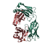
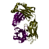
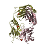
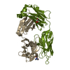
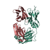
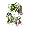
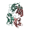
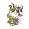
- PDB-3c08: Crystal structure the Fab fragment of matuzumab/EMD72000 (Fab72000) -
+ Open data

- Basic information
| Entry | Database: PDB / ID: 3c08 | ||||||
|---|---|---|---|---|---|---|---|
| Title | Crystal structure the Fab fragment of matuzumab/EMD72000 (Fab72000) | ||||||
 Components Components |
| ||||||
 Keywords Keywords |  IMMUNE SYSTEM / FAB FRAGMENT / ANTITUMOR / DRUG IMMUNE SYSTEM / FAB FRAGMENT / ANTITUMOR / DRUG | ||||||
| Function / homology | Immunoglobulins / Immunoglobulin-like / Sandwich / Mainly Beta Function and homology information Function and homology information | ||||||
| Biological species |  Mus musculus (house mouse) Mus musculus (house mouse) | ||||||
| Method | X-RAY DIFFRACTION / SYNCHROTRON / MOLECULAR REPLACEMENT / molecular replacement / Resolution: 2.15 Å | ||||||
 Authors Authors | Ferguson, K.M. / Schmiedel, J. / Knoechel, T. | ||||||
 Citation Citation | Journal: Cancer Cell / Year: 2008 Title: Matuzumab binding to EGFR prevents the conformational rearrangement required for dimerization. Authors: Schmiedel, J. / Blaukat, A. / Li, S. / Knochel, T. / Ferguson, K.M. | ||||||
| History |
|
- Structure visualization
| Structure viewer | Molecule: MolmilJmol/JSmol |
|---|
- Downloads & links
-Download
| PDBx/mmCIF format | 3c08.cif.gz | 94.6 KB | Display | PDBx/mmCIF format |
|---|---|---|---|---|
| PDB format | pdb3c08.ent.gz | 68.8 KB | Display | PDB format |
| PDBx/mmJSON format | 3c08.json.gz | Tree view | PDBx/mmJSON format | |
| Others |  Other downloads Other downloads |
-Validation report
| Arichive directory | https://data.pdbj.org/pub/pdb/validation_reports/c0/3c08ftp://data.pdbj.org/pub/pdb/validation_reports/c0/3c08 | HTTPS FTP |
|---|
-Related structure data
| Related structure data | 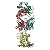 3c09C  1l7iS C: citing same article ( S: Starting model for refinement |
|---|---|
| Similar structure data |


-Links
 PDBj
PDBj

- Assembly
| Deposited unit | 
| ||||||||
|---|---|---|---|---|---|---|---|---|---|
| 1 |
| ||||||||
| Unit cell |
|
-Components
| #1: Antibody | Mass: 23235.678 Da / Num. of mol.: 1 Source method: isolated from a genetically manipulated source Details: Human/mouse chimeric derivative of mouse monoclonal antibody 425 Source: (gene. exp.) Mus musculus (house mouse) / Species: , / Strain: , / Description: humanized mouse / Cell line (production host): MYELOMA / Production host: Mus musculus (house mouse) | ||
|---|---|---|---|
| #2: Antibody | Mass: 24087.777 Da / Num. of mol.: 1 Source method: isolated from a genetically manipulated source Details: Human/mouse chimeric derivative of mouse monoclonal antibody 425 Source: (gene. exp.) Mus musculus (house mouse) / Species: , / Strain: , / Description: humanized mouse / Cell line (production host): MYELOMA / Production host: Mus musculus (house mouse) | ||
| #3: Chemical | Sulfate  Mass: 96.063 Da / Num. of mol.: 2 / Source method: obtained synthetically / Formula: SO4 Mass: 96.063 Da / Num. of mol.: 2 / Source method: obtained synthetically / Formula: SO4#4: Water | ChemComp-HOH / | Water Mass: 18.015 Da / Num. of mol.: 99 / Source method: isolated from a natural source / Formula: H2O Mass: 18.015 Da / Num. of mol.: 99 / Source method: isolated from a natural source / Formula: H2O |
-Experimental details
-Experiment
| Experiment | Method: X-RAY DIFFRACTION / Number of used crystals: 1 |
|---|
- Sample preparation
| Crystal | Density Matthews: 1.79 Å3/Da / Density % sol: 31.34 % |
|---|---|
| Crystal grow | Temperature: 298 K / Method: vapor diffusion / pH: 6.5 Details: 1.8M ammonium sulfate, 0.1M MES, pH 6.5, vapor diffusion, temperature 298K |
-Data collection
| Diffraction | Mean temperature: 100 K |
|---|---|
| Diffraction source | Source: SYNCHROTRON / Site: CHESS  / Beamline: F1 / Wavelength: 0.918 Å / Beamline: F1 / Wavelength: 0.918 Å |
| Detector | Type: ADSC QUANTUM 210 / Detector: CCD / Date: Jan 7, 2007 |
| Radiation | Protocol: SINGLE WAVELENGTH / Monochromatic (M) / Laue (L): M / Scattering type: x-ray |
| Radiation wavelength | Wavelength: 0.918 Å / Relative weight: 1 |
| Reflection | Resolution: 2.15→52.7 Å / Num. all: 20191 / Num. obs: 20191 / % possible obs: 99.9 % / Observed criterion σ(F): 0 / Observed criterion σ(I): 0 / Redundancy: 5.3 % / Rmerge(I) obs: 0.102 / Rsym value: 0.102 / Χ2: 1.361 / Net I/σ(I): 7.2 |
| Reflection shell | Resolution: 2.15→2.23 Å / Redundancy: 4.6 % / Rmerge(I) obs: 0.417 / Mean I/σ(I) obs: 3.618 / Num. unique all: 1971 / Rsym value: 0.417 / Χ2: 0.931 / % possible all: 99.9 |
-Phasing
| Phasing | Method: molecular replacement | ||||||||||||||||||||||||||||||||||||||||||||||||||||||||||||||||||||||||||||||||||||||||||||||||||||||||||||||||
|---|---|---|---|---|---|---|---|---|---|---|---|---|---|---|---|---|---|---|---|---|---|---|---|---|---|---|---|---|---|---|---|---|---|---|---|---|---|---|---|---|---|---|---|---|---|---|---|---|---|---|---|---|---|---|---|---|---|---|---|---|---|---|---|---|---|---|---|---|---|---|---|---|---|---|---|---|---|---|---|---|---|---|---|---|---|---|---|---|---|---|---|---|---|---|---|---|---|---|---|---|---|---|---|---|---|---|---|---|---|---|---|---|---|
| Phasing MR | Model details: Phaser MODE: MR_AUTO
| ||||||||||||||||||||||||||||||||||||||||||||||||||||||||||||||||||||||||||||||||||||||||||||||||||||||||||||||||
| Phasing dm | Method: Solvent flattening and Histogram matching / Reflection: 20127 | ||||||||||||||||||||||||||||||||||||||||||||||||||||||||||||||||||||||||||||||||||||||||||||||||||||||||||||||||
| Phasing dm shell |
|
- Processing
| Software |
| ||||||||||||||||||||||||||||||||||||||||||||||||||||||||||||||||||||||||||||||||||||||||||
|---|---|---|---|---|---|---|---|---|---|---|---|---|---|---|---|---|---|---|---|---|---|---|---|---|---|---|---|---|---|---|---|---|---|---|---|---|---|---|---|---|---|---|---|---|---|---|---|---|---|---|---|---|---|---|---|---|---|---|---|---|---|---|---|---|---|---|---|---|---|---|---|---|---|---|---|---|---|---|---|---|---|---|---|---|---|---|---|---|---|---|---|
| Refinement | Method to determine structure: MOLECULAR REPLACEMENT Starting model: PDB id 1L7I Resolution: 2.15→50 Å / Cor.coef. Fo:Fc: 0.93 / Cor.coef. Fo:Fc free: 0.897 / Cross valid method: THROUGHOUT / σ(F): 0 / σ(I): 0 / ESU R: 0.362 / ESU R Free: 0.239 / Stereochemistry target values: MAXIMUM LIKELIHOOD / Details: HYDROGENS HAVE BEEN ADDED IN THE RIDING POSITIONS
| ||||||||||||||||||||||||||||||||||||||||||||||||||||||||||||||||||||||||||||||||||||||||||
| Solvent computation | Ion probe radii: 0.8 Å / Shrinkage radii: 0.8 Å / VDW probe radii: 1.2 Å / Solvent model: MASK | ||||||||||||||||||||||||||||||||||||||||||||||||||||||||||||||||||||||||||||||||||||||||||
| Displacement parameters | Biso mean: 25.434 Å2
| ||||||||||||||||||||||||||||||||||||||||||||||||||||||||||||||||||||||||||||||||||||||||||
| Refinement step | Cycle: LAST / Resolution: 2.15→50 Å
| ||||||||||||||||||||||||||||||||||||||||||||||||||||||||||||||||||||||||||||||||||||||||||
| Refine LS restraints |
| ||||||||||||||||||||||||||||||||||||||||||||||||||||||||||||||||||||||||||||||||||||||||||
| LS refinement shell | Resolution: 2.145→2.201 Å / Total num. of bins used: 20
|
