 Movie
Movie Controller

[English] 日本語
 Yorodumi
- PDB-3bx9: Monomeric Far-red Fluorescent Protein mKate Crystallized at pH 2.0 -
+ Open data

- Basic information
| Entry | Database: PDB / ID: 3bx9 | ||||||
|---|---|---|---|---|---|---|---|
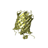
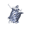
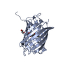
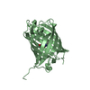
| Title | Monomeric Far-red Fluorescent Protein mKate Crystallized at pH 2.0 | ||||||
 Components Components | Far-red fluorescent protein mKate | ||||||
 Keywords Keywords |  FLUORESCENT PROTEIN / Far-red fluorescent protein / E. quadricolor / Chromophore structure / pH-induced cis-trans izomerization FLUORESCENT PROTEIN / Far-red fluorescent protein / E. quadricolor / Chromophore structure / pH-induced cis-trans izomerization | ||||||
| Function / homology | Green Fluorescent Protein / Green fluorescent protein / Beta Barrel / Mainly Beta / CITRIC ACID Function and homology information Function and homology information | ||||||
| Biological species |  Entacmaea quadricolor (sea anemone) Entacmaea quadricolor (sea anemone) | ||||||
| Method | X-RAY DIFFRACTION / SYNCHROTRON / MOLECULAR REPLACEMENT / Resolution: 1.8 Å | ||||||
 Authors Authors | Pletnev, S. / Pletneva, N. / Pletnev, V. | ||||||
 Citation Citation | Journal: J.Biol.Chem. / Year: 2008 Title: A Crystallographic Study of Bright Far-Red Fluorescent Protein mKate Reveals pH-induced cis-trans Isomerization of the Chromophore. Authors: Pletnev, S. / Shcherbo, D. / Chudakov, D.M. / Pletneva, N. / Merzlyak, E.M. / Wlodawer, A. / Dauter, Z. / Pletnev, V. | ||||||
| History |
|
- Structure visualization
| Structure viewer | Molecule: MolmilJmol/JSmol |
|---|
- Downloads & links
-Download
| PDBx/mmCIF format | 3bx9.cif.gz | 111.5 KB | Display | PDBx/mmCIF format |
|---|---|---|---|---|
| PDB format | pdb3bx9.ent.gz | 85.4 KB | Display | PDB format |
| PDBx/mmJSON format | 3bx9.json.gz | Tree view | PDBx/mmJSON format | |
| Others |  Other downloads Other downloads |
-Validation report
| Arichive directory | https://data.pdbj.org/pub/pdb/validation_reports/bx/3bx9ftp://data.pdbj.org/pub/pdb/validation_reports/bx/3bx9 | HTTPS FTP |
|---|
-Related structure data
| Related structure data |  3bxaC  3bxbC  3bxcC  1uisS S: Starting model for refinement C: citing same article ( |
|---|---|
| Similar structure data |


-Links
 PDBj
PDBj- Assembly
| Deposited unit | 
| ||||||||
|---|---|---|---|---|---|---|---|---|---|
| 1 | 
| ||||||||
| 2 | 
| ||||||||
| 3 | 
| ||||||||
| Unit cell |
|
-Components
| #1: Protein | Mass: 27640.482 Da / Num. of mol.: 2 Source method: isolated from a genetically manipulated source Source: (gene. exp.) Entacmaea quadricolor (sea anemone) / Production host:  Escherichia coli (E. coli) / Strain (production host): XL1 Blue Escherichia coli (E. coli) / Strain (production host): XL1 Blue#2: Chemical | ChemComp-CIT / Citric acid  Mass: 192.124 Da / Num. of mol.: 6 / Source method: obtained synthetically / Formula: C6H8O7 Mass: 192.124 Da / Num. of mol.: 6 / Source method: obtained synthetically / Formula: C6H8O7#3: Chemical | Glycerol  Mass: 92.094 Da / Num. of mol.: 2 / Source method: obtained synthetically / Formula: C3H8O3 Mass: 92.094 Da / Num. of mol.: 2 / Source method: obtained synthetically / Formula: C3H8O3#4: Water | ChemComp-HOH / | Water Mass: 18.015 Da / Num. of mol.: 259 / Source method: isolated from a natural source / Formula: H2O Mass: 18.015 Da / Num. of mol.: 259 / Source method: isolated from a natural source / Formula: H2O |
|---|
-Experimental details
-Experiment
| Experiment | Method: X-RAY DIFFRACTION / Number of used crystals: 1 |
|---|
- Sample preparation
| Crystal | Density Matthews: 2.62 Å3/Da / Density % sol: 52.97 % |
|---|---|
| Crystal grow | Temperature: 293 K / pH: 2 Details: 20% w/v PEG 3350, 0.2M ammonium citrate tribasic, 0.4M citric acid, pH 2.0, VAPOR DIFFUSION, HANGING DROP, temperature 293K |
-Data collection
| Diffraction | Mean temperature: 100 K |
|---|---|
| Diffraction source | Source: SYNCHROTRON / Site: APS  / Beamline: 22-ID / Wavelength: 1 Å / Beamline: 22-ID / Wavelength: 1 Å |
| Detector | Type: MARMOSAIC 300 mm CCD / Detector: CCD |
| Radiation | Protocol: SINGLE WAVELENGTH / Monochromatic (M) / Laue (L): M / Scattering type: x-ray |
| Radiation wavelength | Wavelength: 1 Å / Relative weight: 1 |
| Reflection | Resolution: 1.8→30.43 Å / Num. all: 53976 / Num. obs: 52642 / % possible obs: 99.9 % / Redundancy: 14.1 % / Rmerge(I) obs: 0.061 |
| Reflection shell | Resolution: 1.8→1.86 Å / Redundancy: 14.4 % / Rmerge(I) obs: 0.556 / Mean I/σ(I) obs: 5.63 / % possible all: 99.7 |
- Processing
| Software |
| ||||||||||||||||||||||||||||||||||||||||||||||||||||||||||||||||||||||||||||||||||||||||||||||||||||||||||||||||||||||||||||||||||||||||||||||||||||||||||||||||||||||||||
|---|---|---|---|---|---|---|---|---|---|---|---|---|---|---|---|---|---|---|---|---|---|---|---|---|---|---|---|---|---|---|---|---|---|---|---|---|---|---|---|---|---|---|---|---|---|---|---|---|---|---|---|---|---|---|---|---|---|---|---|---|---|---|---|---|---|---|---|---|---|---|---|---|---|---|---|---|---|---|---|---|---|---|---|---|---|---|---|---|---|---|---|---|---|---|---|---|---|---|---|---|---|---|---|---|---|---|---|---|---|---|---|---|---|---|---|---|---|---|---|---|---|---|---|---|---|---|---|---|---|---|---|---|---|---|---|---|---|---|---|---|---|---|---|---|---|---|---|---|---|---|---|---|---|---|---|---|---|---|---|---|---|---|---|---|---|---|---|---|---|---|---|
| Refinement | Method to determine structure: MOLECULAR REPLACEMENT Starting model: PDB ENTRY 1UIS Resolution: 1.8→30 Å / Cor.coef. Fo:Fc: 0.955 / Cor.coef. Fo:Fc free: 0.932 / SU B: 3.224 / SU ML: 0.099 / Stereochemistry target values: MAXIMUM LIKELIHOOD / Details: HYDROGENS HAVE BEEN ADDED IN THE RIDING POSITIONS
| ||||||||||||||||||||||||||||||||||||||||||||||||||||||||||||||||||||||||||||||||||||||||||||||||||||||||||||||||||||||||||||||||||||||||||||||||||||||||||||||||||||||||||
| Solvent computation | Solvent model: MASK | ||||||||||||||||||||||||||||||||||||||||||||||||||||||||||||||||||||||||||||||||||||||||||||||||||||||||||||||||||||||||||||||||||||||||||||||||||||||||||||||||||||||||||
| Displacement parameters | Biso mean: 27.12 Å2
| ||||||||||||||||||||||||||||||||||||||||||||||||||||||||||||||||||||||||||||||||||||||||||||||||||||||||||||||||||||||||||||||||||||||||||||||||||||||||||||||||||||||||||
| Refinement step | Cycle: LAST / Resolution: 1.8→30 Å
| ||||||||||||||||||||||||||||||||||||||||||||||||||||||||||||||||||||||||||||||||||||||||||||||||||||||||||||||||||||||||||||||||||||||||||||||||||||||||||||||||||||||||||
| Refine LS restraints |
| ||||||||||||||||||||||||||||||||||||||||||||||||||||||||||||||||||||||||||||||||||||||||||||||||||||||||||||||||||||||||||||||||||||||||||||||||||||||||||||||||||||||||||
| LS refinement shell | Resolution: 1.8→1.85 Å
|
