 Movie
Movie Controller

[English] 日本語
 Yorodumi
- PDB-3bw4: Crystal structures and site-directed mutagenesis study of nitroal... -
+ Open data

- Basic information
| Entry | Database: PDB / ID: 3bw4 | ||||||
|---|---|---|---|---|---|---|---|
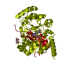
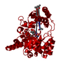
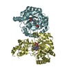
| Title | Crystal structures and site-directed mutagenesis study of nitroalkane oxidase from Streptomyces ansochromogenes | ||||||
 Components Components | 2-nitropropane dioxygenase | ||||||
 Keywords Keywords | OXIDOREDUCTASE / TIM barrel / Dioxygenase | ||||||
| Function / homology |  Function and homology informationnitronate monooxygenase activity / dioxygenase activity / response to toxic substance / nucleotide binding Function and homology informationnitronate monooxygenase activity / dioxygenase activity / response to toxic substance / nucleotide bindingSimilarity search - Function | ||||||
| Biological species |  Streptomyces ansochromogenes (bacteria) Streptomyces ansochromogenes (bacteria) | ||||||
| Method | X-RAY DIFFRACTION / MOLECULAR REPLACEMENT / Resolution: 2.2 Å | ||||||
 Authors Authors | Li, Y.H. / Gao, Z.Q. / Hou, H.F. | ||||||
 Citation Citation | Journal: To be Published Title: Crystal structures and site-directed mutagenesis study of nitroalkane oxidase from Streptomyces ansochromogenes Authors: Li, Y.H. / Gao, Z.Q. / Hou, H.F. / Dong, Y.H. / Tan, H.R. | ||||||
| History |
|
- Structure visualization
| Structure viewer | Molecule: MolmilJmol/JSmol |
|---|
- Downloads & links
-Download
| PDBx/mmCIF format | 3bw4.cif.gz | 80.6 KB | Display | PDBx/mmCIF format |
|---|---|---|---|---|
| PDB format | pdb3bw4.ent.gz | 59.2 KB | Display | PDB format |
| PDBx/mmJSON format | 3bw4.json.gz | Tree view | PDBx/mmJSON format | |
| Others |  Other downloads Other downloads |
-Validation report
| Arichive directory | https://data.pdbj.org/pub/pdb/validation_reports/bw/3bw4ftp://data.pdbj.org/pub/pdb/validation_reports/bw/3bw4 | HTTPS FTP |
|---|
-Related structure data
| Related structure data |  3bw2S S: Starting model for refinement |
|---|---|
| Similar structure data |


-Links
 PDBj
PDBj- Assembly
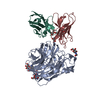
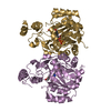
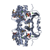
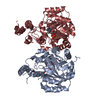
| Deposited unit | 
| ||||||||
|---|---|---|---|---|---|---|---|---|---|
| 1 | 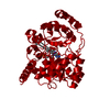
| ||||||||
| Unit cell |
|
-Components
| #1: Protein | Mass: 38521.328 Da / Num. of mol.: 1 / Mutation: W320Y Source method: isolated from a genetically manipulated source Source: (gene. exp.) Streptomyces ansochromogenes (bacteria)Strain: 7100 / Gene: 2-npdl / Plasmid: pET23b / Production host: Escherichia coli (E. coli) / Strain (production host): BL21(DE3) / References: UniProt: Q9FDD4, nitroalkane oxidase |
|---|---|
| #2: Chemical | ChemComp-FMN / Flavin mononucleotide  Mass: 456.344 Da / Num. of mol.: 1 / Source method: obtained synthetically / Formula: C17H21N4O9P Mass: 456.344 Da / Num. of mol.: 1 / Source method: obtained synthetically / Formula: C17H21N4O9P |
| #3: Chemical | ChemComp-MPD / (2-Methyl-2,4-pentanediol  Mass: 118.174 Da / Num. of mol.: 1 / Source method: obtained synthetically / Formula: C6H14O2 / Comment: precipitant*YM Mass: 118.174 Da / Num. of mol.: 1 / Source method: obtained synthetically / Formula: C6H14O2 / Comment: precipitant*YM |
| #4: Water | ChemComp-HOH / Water Mass: 18.015 Da / Num. of mol.: 136 / Source method: isolated from a natural source / Formula: H2O Mass: 18.015 Da / Num. of mol.: 136 / Source method: isolated from a natural source / Formula: H2O |
-Experimental details
-Experiment
| Experiment | Method: X-RAY DIFFRACTION / Number of used crystals: 1 |
|---|
- Sample preparation
| Crystal | Density Matthews: 2.4 Å3/Da / Density % sol: 48.81 % |
|---|---|
| Crystal grow | Temperature: 289 K / Method: vapor diffusion, hanging drop / pH: 8.5 Details: 0.2M ammonium dihydrogen phosphate, 0.1M Tris-Cl, 50% MPD, pH8.5, VAPOR DIFFUSION, HANGING DROP, temperature 289K |
-Data collection
| Diffraction | Mean temperature: 100 K |
|---|---|
| Diffraction source | Source: ROTATING ANODE / Type: RIGAKU MICROMAX-007 / Wavelength: 1.5418 Å |
| Detector | Type: MAR scanner 345 mm plate / Detector: IMAGE PLATE / Date: Jul 15, 2007 |
| Radiation | Protocol: SINGLE WAVELENGTH / Monochromatic (M) / Laue (L): M / Scattering type: x-ray |
| Radiation wavelength | Wavelength: 1.5418 Å / Relative weight: 1 |
| Reflection | Resolution: 2→30 Å / Num. all: 25547 / Num. obs: 22020 / % possible obs: 86.2 % / Observed criterion σ(F): 1 / Observed criterion σ(I): 1 / Redundancy: 9.8 % / Rmerge(I) obs: 0.089 |
| Reflection shell | Resolution: 2→2.07 Å / Redundancy: 9.1 % / Rmerge(I) obs: 0.53 / Num. unique all: 2524 / % possible all: 99.6 |
- Processing
| Software |
| |||||||||||||||||||||||||
|---|---|---|---|---|---|---|---|---|---|---|---|---|---|---|---|---|---|---|---|---|---|---|---|---|---|---|
| Refinement | Method to determine structure: MOLECULAR REPLACEMENT Starting model: PDB ENTRY 3BW2 Resolution: 2.2→30 Å / σ(F): 1 / Stereochemistry target values: maximum likelihood
| |||||||||||||||||||||||||
| Displacement parameters | Biso mean: 29.12 Å2
| |||||||||||||||||||||||||
| Refinement step | Cycle: LAST / Resolution: 2.2→30 Å
| |||||||||||||||||||||||||
| Refine LS restraints |
| |||||||||||||||||||||||||
| Xplor file |
|
