 Movie
Movie Controller

[English] 日本語
 Yorodumi
- PDB-3bsz: Crystal structure of the transthyretin-retinol binding protein-Fa... -
+ Open data

- Basic information
| Entry | Database: PDB / ID: 3bsz | ||||||
|---|---|---|---|---|---|---|---|
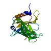
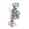
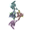
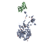
| Title | Crystal structure of the transthyretin-retinol binding protein-Fab complex | ||||||
 Components Components |
| ||||||
 Keywords Keywords | TRANSPORT PROTEIN/IMMUNE SYSTEM /  retinol / Vitamin A / protein-protein complex / RBP / TTR / Amyloid / Disease mutation / Glycoprotein / Hormone / Polyneuropathy / Retinol-binding / Secreted / Thyroid hormone / Transport / Sensory transduction / Vision / TRANSPORT PROTEIN-IMMUNE SYSTEM COMPLEX retinol / Vitamin A / protein-protein complex / RBP / TTR / Amyloid / Disease mutation / Glycoprotein / Hormone / Polyneuropathy / Retinol-binding / Secreted / Thyroid hormone / Transport / Sensory transduction / Vision / TRANSPORT PROTEIN-IMMUNE SYSTEM COMPLEX | ||||||
| Function / homology |  Function and homology information Function and homology informationRetinoid metabolism disease events / urinary bladder development / embryonic retina morphogenesis in camera-type eye / retinol transport / female genitalia morphogenesis / retinol transmembrane transporter activity / embryonic organ morphogenesis / maintenance of gastrointestinal epithelium / embryonic skeletal system development / negative regulation of cardiac muscle cell proliferation ...Retinoid metabolism disease events / urinary bladder development / embryonic retina morphogenesis in camera-type eye / retinol transport / female genitalia morphogenesis / retinol transmembrane transporter activity / embryonic organ morphogenesis / maintenance of gastrointestinal epithelium / embryonic skeletal system development / negative regulation of cardiac muscle cell proliferation / eye development / heart trabecula formation / cardiac muscle tissue development / retinal binding / retinol metabolic process / retinol binding / positive regulation of immunoglobulin production / Retinoid cycle disease events / The canonical retinoid cycle in rods (twilight vision) / thyroid hormone binding / uterus development / vagina development / purine nucleobase metabolic process / Non-integrin membrane-ECM interactions / response to retinoic acid / Retinoid metabolism and transport / visual perception / gluconeogenesis / lung development / positive regulation of insulin secretion / hormone activity / azurophil granule lumen / glucose homeostasis / heart development / Amyloid fiber formation / Neutrophil degranulation / extracellular space / extracellular exosome / extracellular region / identical protein bindingSimilarity search - Function | ||||||
| Biological species |  Homo sapiens (human) Homo sapiens (human) Mus musculus (house mouse) Mus musculus (house mouse) | ||||||
| Method | X-RAY DIFFRACTION / SYNCHROTRON / MOLECULAR REPLACEMENT / Resolution: 3.38 Å | ||||||
 Authors Authors | Zanotti, G. / Cendron, L. / Gliubich, F. / Folli, C. / Berni, R. | ||||||
 Citation Citation | Journal: Febs J. / Year: 2008 Title: Structural and mutational analyses of protein-protein interactions between transthyretin and retinol-binding protein. Authors: Zanotti, G. / Folli, C. / Cendron, L. / Alfieri, B. / Nishida, S.K. / Gliubich, F. / Pasquato, N. / Negro, A. / Berni, R. #1: Journal: Science / Year: 1995Title: Structure of a complex of two plasma proteins: transthyretin and retinol-binding protein. Authors: Monaco, H.L. / Rizzi, M. / Coda, A. #2: Journal: Biochemistry / Year: 1999Title: The structure of human retinol-binding protein (RBP) with its carrier protein transthyretin reveals an interaction with the carboxy terminus of RBP. Authors: Naylor, H.M. / Newcomer, M.E. #3: Journal: ACTA CRYSTALLOGR.,SECT.D / Year: 1999 Title: Crystallization and preliminary X-ray data for the human transthyretin-retinol-binding protein (RBP) complex bound to an anti-RBP Fab. Authors: Malpeli, G. / Zanotti, G. / Gliubich, F. / Rizzotto, A. / Nishida, S.K. / Folli, C. / Berni, R. | ||||||
| History |
|
- Structure visualization
| Structure viewer | Molecule: MolmilJmol/JSmol |
|---|
- Downloads & links
-Download
| PDBx/mmCIF format | 3bsz.cif.gz | 336.7 KB | Display | PDBx/mmCIF format |
|---|---|---|---|---|
| PDB format | pdb3bsz.ent.gz | 271.5 KB | Display | PDB format |
| PDBx/mmJSON format | 3bsz.json.gz | Tree view | PDBx/mmJSON format | |
| Others |  Other downloads Other downloads |
-Validation report
| Arichive directory | https://data.pdbj.org/pub/pdb/validation_reports/bs/3bszftp://data.pdbj.org/pub/pdb/validation_reports/bs/3bsz | HTTPS FTP |
|---|
-Related structure data
| Related structure data |  3bt0C  3cxfC 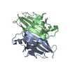 1f41S 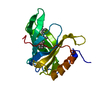 1rbpS S: Starting model for refinement C: citing same article ( |
|---|---|
| Similar structure data |


-Links
 PDBj
PDBj


- Assembly
| Deposited unit | 
| ||||||||
|---|---|---|---|---|---|---|---|---|---|
| 1 |
| ||||||||
| Unit cell |
|
-Components
-Protein , 2 types, 6 molecules ABCDEF
| #1: Protein | / Prealbumin / TBPA / TTR / ATTR Mass: 13777.360 Da / Num. of mol.: 4 Source method: isolated from a genetically manipulated source Source: (gene. exp.) Homo sapiens (human) / Gene: TTR, PALB / Plasmid: pET11B-hTTR / Production host:  Escherichia coli (E. coli) / Strain (production host): BL21(DE3) / References: UniProt: P02766 Escherichia coli (E. coli) / Strain (production host): BL21(DE3) / References: UniProt: P02766#2: Protein | Mass: 20226.605 Da / Num. of mol.: 2 / Source method: isolated from a natural source / Source: (natural) Homo sapiens (human) / Secretion: Plasma / References: UniProt: P02753 |
|---|
-Antibody , 2 types, 4 molecules LMHN
| #3: Antibody | Fragment antigen-binding Mass: 23708.055 Da / Num. of mol.: 2 Source method: isolated from a genetically manipulated source Source: (gene. exp.) Mus musculus (house mouse) / Gene: A8P3 MAb#4: Antibody | Fragment antigen-bindingMass: 22939.566 Da / Num. of mol.: 2 Source method: isolated from a genetically manipulated source Source: (gene. exp.) Mus musculus (house mouse) / Gene: A8P3 MAb |
|---|
-Non-polymers , 2 types, 336 molecules 

| #5: Chemical | Retinol Mass: 286.452 Da / Num. of mol.: 2 / Source method: obtained synthetically / Formula: C20H30O Mass: 286.452 Da / Num. of mol.: 2 / Source method: obtained synthetically / Formula: C20H30O#6: Water | ChemComp-HOH / | WaterMass: 18.015 Da / Num. of mol.: 334 / Source method: isolated from a natural source / Formula: H2O |
|---|
-Experimental details
-Experiment
| Experiment | Method: X-RAY DIFFRACTION / Number of used crystals: 2 |
|---|
- Sample preparation
| Crystal | Density Matthews: 2.86 Å3/Da / Density % sol: 57.04 % |
|---|---|
| Crystal grow | Temperature: 295 K / Method: vapor diffusion, sitting drop / pH: 5 Details: 2.35M ammonium phosphate, 10mM sodium citrate, 10mM beta-mercaptoethanol, pH 5.0, VAPOR DIFFUSION, SITTING DROP, temperature 295K |
-Data collection
| Diffraction | Mean temperature: 100 K |
|---|---|
| Diffraction source | Source: SYNCHROTRON / Site: ELETTRA  / Beamline: 5.2R / Wavelength: 1.3 Å / Beamline: 5.2R / Wavelength: 1.3 Å |
| Detector | Type: MAR scanner 180 mm plate / Detector: IMAGE PLATE / Date: Oct 21, 1997 |
| Radiation | Monochromator: Si (111) / Protocol: SINGLE WAVELENGTH / Monochromatic (M) / Laue (L): M / Scattering type: x-ray |
| Radiation wavelength | Wavelength: 1.3 Å / Relative weight: 1 |
| Reflection | Resolution: 3.36→55 Å / Num. all: 25746 / Num. obs: 25746 / % possible obs: 84.4 % / Observed criterion σ(F): 0 / Observed criterion σ(I): 0 / Redundancy: 3 % / Rmerge(I) obs: 0.16 / Net I/σ(I): 3.3 |
| Reflection shell | Resolution: 3.36→3.51 Å / Redundancy: 2.6 % / Rmerge(I) obs: 0.34 / Mean I/σ(I) obs: 1.9 / Num. unique all: 1622 / % possible all: 77.7 |
- Processing
| Software |
| ||||||||||||||||||||||||||||||||||||||||||||||||||||||||||||
|---|---|---|---|---|---|---|---|---|---|---|---|---|---|---|---|---|---|---|---|---|---|---|---|---|---|---|---|---|---|---|---|---|---|---|---|---|---|---|---|---|---|---|---|---|---|---|---|---|---|---|---|---|---|---|---|---|---|---|---|---|---|
| Refinement | Method to determine structure: MOLECULAR REPLACEMENT Starting model: 1RBP, 1F41 Resolution: 3.38→15.72 Å / Rfactor Rfree error: 0.009 / Data cutoff high absF: 8281753.41 / Data cutoff low absF: 0 / Isotropic thermal model: RESTRAINED / Cross valid method: THROUGHOUT / σ(F): 0 / σ(I): 0 / Stereochemistry target values: Engh & Huber
| ||||||||||||||||||||||||||||||||||||||||||||||||||||||||||||
| Solvent computation | Solvent model: FLAT MODEL / Bsol: 48.9765 Å2 / ksol: 0.32803 e/Å3 | ||||||||||||||||||||||||||||||||||||||||||||||||||||||||||||
| Displacement parameters | Biso mean: 37.5 Å2
| ||||||||||||||||||||||||||||||||||||||||||||||||||||||||||||
| Refine analyze |
| ||||||||||||||||||||||||||||||||||||||||||||||||||||||||||||
| Refinement step | Cycle: LAST / Resolution: 3.38→15.72 Å
| ||||||||||||||||||||||||||||||||||||||||||||||||||||||||||||
| Refine LS restraints |
| ||||||||||||||||||||||||||||||||||||||||||||||||||||||||||||
| Refine LS restraints NCS | NCS model details: CONSTR | ||||||||||||||||||||||||||||||||||||||||||||||||||||||||||||
| LS refinement shell | Resolution: 3.36→3.57 Å / Rfactor Rfree error: 0.034 / Total num. of bins used: 6
| ||||||||||||||||||||||||||||||||||||||||||||||||||||||||||||
| Xplor file |
|
