 Movie
Movie Controller


+ Open data

- Basic information
| Entry | Database: PDB / ID: 3bqp | ||||||
|---|---|---|---|---|---|---|---|
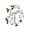
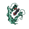
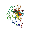
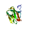
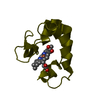
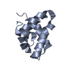
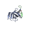
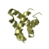
| Title | Crystal Structure of Human Saposin D (orthorhombic) | ||||||
 Components Components | Proactivator polypeptide | ||||||
 Keywords Keywords | LIPID BINDING PROTEIN /  SAPOSIN / SPHINGOLIPID ACTIVATOR PROTEIN / ACID CERAMIDASE / FARBER DISEASE / LIPID METABOLISM / LYSOSOME / SPHINGOLIPID METABOLISM SAPOSIN / SPHINGOLIPID ACTIVATOR PROTEIN / ACID CERAMIDASE / FARBER DISEASE / LIPID METABOLISM / LYSOSOME / SPHINGOLIPID METABOLISM | ||||||
| Function / homology |  Function and homology information Function and homology informationpositive regulation of beta-galactosidase activity / ganglioside GM1 transport to membrane / ganglioside GM2 binding / ganglioside GM3 binding / ganglioside GP1c binding / ganglioside GM1 binding / ganglioside GT1b binding / prostate gland growth / sphingolipid metabolic process / Glycosphingolipid catabolism ...positive regulation of beta-galactosidase activity / ganglioside GM1 transport to membrane / ganglioside GM2 binding / ganglioside GM3 binding / ganglioside GP1c binding / ganglioside GM1 binding / ganglioside GT1b binding / prostate gland growth / sphingolipid metabolic process / Glycosphingolipid catabolism / epithelial cell differentiation involved in prostate gland development / lysosomal transport / azurophil granule membrane / regulation of lipid metabolic process / enzyme activator activity / adenylate cyclase-inhibiting G protein-coupled receptor signaling pathway / lysosomal lumen / Peptide ligand-binding receptors / regulation of autophagy / phospholipid binding / late endosome / Platelet degranulation / G alpha (i) signalling events / scaffold protein binding / collagen-containing extracellular matrix / protease binding / lysosome / lysosomal membrane / intracellular membrane-bounded organelle / Neutrophil degranulation / protein homodimerization activity / extracellular space / extracellular exosome / extracellular region / identical protein binding / plasma membraneSimilarity search - Function | ||||||
| Biological species |  Homo sapiens (human) Homo sapiens (human) | ||||||
| Method | X-RAY DIFFRACTION / SYNCHROTRON / SAD / Resolution: 1.3 Å | ||||||
 Authors Authors | Prive, G.G. / Popovic, K. | ||||||
 Citation Citation | Journal: Acta Crystallogr.,Sect.D / Year: 2008 Title: Structures of the human ceramide activator protein saposin D. Authors: Popovic, K. / Prive, G.G. | ||||||
| History |
|
- Structure visualization
| Structure viewer | Molecule: MolmilJmol/JSmol |
|---|
- Downloads & links
-Download
| PDBx/mmCIF format | 3bqp.cif.gz | 81 KB | Display | PDBx/mmCIF format |
|---|---|---|---|---|
| PDB format | pdb3bqp.ent.gz | 65.5 KB | Display | PDB format |
| PDBx/mmJSON format | 3bqp.json.gz | Tree view | PDBx/mmJSON format | |
| Others |  Other downloads Other downloads |
-Validation report
| Arichive directory | https://data.pdbj.org/pub/pdb/validation_reports/bq/3bqpftp://data.pdbj.org/pub/pdb/validation_reports/bq/3bqp | HTTPS FTP |
|---|
-Related structure data


-Links
 PDBj
PDBj


- Assembly
| Deposited unit | 
| ||||||||
|---|---|---|---|---|---|---|---|---|---|
| 1 | 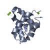
| ||||||||
| 2 | 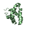
| ||||||||
| Unit cell |
|
-Components
| #1: Protein | Mass: 8926.332 Da / Num. of mol.: 2 / Fragment: SAPOSIN D, RESIDUES 405-484 Source method: isolated from a genetically manipulated source Source: (gene. exp.) Homo sapiens (human) / Gene: PSAP; / Plasmid: PET-16 / Production host:  Escherichia coli (E. coli) / Strain (production host): BL21(DE3) ROSETTA-GAMI 2 / References: UniProt: P07602 Escherichia coli (E. coli) / Strain (production host): BL21(DE3) ROSETTA-GAMI 2 / References: UniProt: P07602#2: Chemical |   Mass: 24.305 Da / Num. of mol.: 3 / Source method: obtained synthetically / Formula: Mg Mass: 24.305 Da / Num. of mol.: 3 / Source method: obtained synthetically / Formula: Mg#3: Water | ChemComp-HOH / | Water Mass: 18.015 Da / Num. of mol.: 198 / Source method: isolated from a natural source / Formula: H2O Mass: 18.015 Da / Num. of mol.: 198 / Source method: isolated from a natural source / Formula: H2O |
|---|
-Experimental details
-Experiment
| Experiment | Method: X-RAY DIFFRACTION / Number of used crystals: 1 |
|---|
- Sample preparation
| Crystal | Density Matthews: 2.25 Å3/Da / Density % sol: 45.27 % |
|---|---|
| Crystal grow | Temperature: 298 K / Method: vapor diffusion, hanging drop / pH: 8.5 Details: PEG 8K, MAGNESIUM CHLORIDE, TRIS.HCL BUFFER, PH 8.5, VAPOR DIFFUSION, HANGING DROP, temperature 298.0K |
-Data collection
| Diffraction | Mean temperature: 100 K |
|---|---|
| Diffraction source | Source: SYNCHROTRON / Site: APS  / Beamline: 19-ID / Wavelength: 1.033 Å / Beamline: 19-ID / Wavelength: 1.033 Å |
| Detector | Type: ADSC QUANTUM 315 / Detector: CCD / Date: Mar 24, 2007 |
| Radiation | Protocol: SINGLE WAVELENGTH / Monochromatic (M) / Laue (L): M / Scattering type: x-ray |
| Radiation wavelength | Wavelength: 1.033 Å / Relative weight: 1 |
| Reflection | Resolution: 1.3→45 Å / Num. obs: 38323 / % possible obs: 99.1 % / Redundancy: 7.5 % / Rmerge(I) obs: 0.044 / Net I/σ(I): 35.4 |
| Reflection shell | Resolution: 1.3→1.35 Å / Redundancy: 6.5 % / Rmerge(I) obs: 0.218 / Mean I/σ(I) obs: 8.1 / % possible all: 98.1 |
- Processing
| Software |
| |||||||||||||||||||||||||||||||||
|---|---|---|---|---|---|---|---|---|---|---|---|---|---|---|---|---|---|---|---|---|---|---|---|---|---|---|---|---|---|---|---|---|---|---|
| Refinement | Method to determine structure: SAD / Resolution: 1.3→34.28 Å / Num. parameters: 13278 / Num. restraintsaints: 16744 / Cross valid method: FREE R / σ(F): 0 / Stereochemistry target values: Engh & Huber / Details: ANISOTROPIC REFINEMENT REDUCED FREE R (NO CUTOFF)
| |||||||||||||||||||||||||||||||||
| Refine analyze | Num. disordered residues: 11 / Occupancy sum hydrogen: 0 / Occupancy sum non hydrogen: 1417 | |||||||||||||||||||||||||||||||||
| Refinement step | Cycle: LAST / Resolution: 1.3→34.28 Å
| |||||||||||||||||||||||||||||||||
| Refine LS restraints |
|
