 Movie
Movie Controller


+ Open data

- Basic information
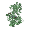
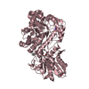
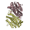
| Entry | Database: PDB / ID: 3bmx | ||||||
|---|---|---|---|---|---|---|---|
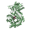
| Title | Beta-N-hexosaminidase (YbbD) from Bacillus subtilis | ||||||
 Components Components | Uncharacterized lipoprotein ybbD | ||||||
 Keywords Keywords |  HYDROLASE / beta-N-hexosaminidase / Bacillus subtilis / TIM barrel / Glycosidase / Lipoprotein / Membrane / Palmitate HYDROLASE / beta-N-hexosaminidase / Bacillus subtilis / TIM barrel / Glycosidase / Lipoprotein / Membrane / Palmitate | ||||||
| Function / homology |  Function and homology informationbeta-N-acetylhexosaminidase activity / beta-N-acetylhexosaminidase / peptidoglycan turnover / N-acetyl-beta-D-galactosaminidase activity / peptidoglycan biosynthetic process / cell wall organization / regulation of cell shape / carbohydrate metabolic process / extracellular region / plasma membrane Function and homology informationbeta-N-acetylhexosaminidase activity / beta-N-acetylhexosaminidase / peptidoglycan turnover / N-acetyl-beta-D-galactosaminidase activity / peptidoglycan biosynthetic process / cell wall organization / regulation of cell shape / carbohydrate metabolic process / extracellular region / plasma membraneSimilarity search - Function | ||||||
| Biological species |  Bacillus subtilis (bacteria) Bacillus subtilis (bacteria) | ||||||
| Method | X-RAY DIFFRACTION / SYNCHROTRON / MOLECULAR REPLACEMENT / Resolution: 1.4 Å | ||||||
 Authors Authors | Fischer, S. | ||||||
 Citation Citation | Journal: J.Biol.Chem. / Year: 2010 Title: Structural and kinetic analysis of Bacillus subtilis N-acetylglucosaminidase reveals a unique Asp-His dyad mechanism Authors: Litzinger, S. / Fischer, S. / Polzer, P. / Diederichs, K. / Welte, W. / Mayer, C. | ||||||
| History |
|
- Structure visualization
| Structure viewer | Molecule: MolmilJmol/JSmol |
|---|
- Downloads & links
-Download
| PDBx/mmCIF format | 3bmx.cif.gz | 540.9 KB | Display | PDBx/mmCIF format |
|---|---|---|---|---|
| PDB format | pdb3bmx.ent.gz | 445.7 KB | Display | PDB format |
| PDBx/mmJSON format | 3bmx.json.gz | Tree view | PDBx/mmJSON format | |
| Others |  Other downloads Other downloads |
-Validation report
| Arichive directory | https://data.pdbj.org/pub/pdb/validation_reports/bm/3bmxftp://data.pdbj.org/pub/pdb/validation_reports/bm/3bmx | HTTPS FTP |
|---|
-Related structure data
| Related structure data |  3nvdC 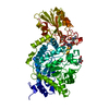 1ex1S  1iix  1j8vS  1lq2S  1x38S S: Starting model for refinement C: citing same article ( |
|---|---|
| Similar structure data |


-Links
 PDBj
PDBj


- Assembly
| Deposited unit | 
| ||||||||
|---|---|---|---|---|---|---|---|---|---|
| 1 | 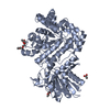
| ||||||||
| 2 | 
| ||||||||
| Unit cell |
|
-Components
| #1: Protein | Mass: 70674.328 Da / Num. of mol.: 2 Source method: isolated from a genetically manipulated source Source: (gene. exp.) Bacillus subtilis (bacteria) / Strain: wt168 / Gene: ybbD / Plasmid: PET16b / Production host: Escherichia coli (E. coli) / Strain (production host): BL21(DE3) / References: UniProt: P40406#2: Chemical | ChemComp-ACT / Acetate  Mass: 59.044 Da / Num. of mol.: 7 / Source method: obtained synthetically / Formula: C2H3O2 Mass: 59.044 Da / Num. of mol.: 7 / Source method: obtained synthetically / Formula: C2H3O2#3: Chemical | ChemComp-NA /   Mass: 22.990 Da / Num. of mol.: 4 / Source method: obtained synthetically / Formula: Na Mass: 22.990 Da / Num. of mol.: 4 / Source method: obtained synthetically / Formula: Na#4: Chemical | Diethylene glycol diethyl ether  Mass: 162.227 Da / Num. of mol.: 3 / Source method: obtained synthetically / Formula: C8H18O3 Mass: 162.227 Da / Num. of mol.: 3 / Source method: obtained synthetically / Formula: C8H18O3#5: Water | ChemComp-HOH / | Water Mass: 18.015 Da / Num. of mol.: 1388 / Source method: isolated from a natural source / Formula: H2O Mass: 18.015 Da / Num. of mol.: 1388 / Source method: isolated from a natural source / Formula: H2O |
|---|
-Experimental details
-Experiment
| Experiment | Method: X-RAY DIFFRACTION / Number of used crystals: 4 |
|---|
- Sample preparation
| Crystal | Density Matthews: 2.37 Å3/Da / Density % sol: 48.13 % |
|---|---|
| Crystal grow | Temperature: 291 K / Method: vapor diffusion, hanging drop / pH: 4.9 Details: 0.1M sodium acetate, 30-33 % PEG 1000, pH 4.9, VAPOR DIFFUSION, HANGING DROP, temperature 291K |
-Data collection
| Diffraction | Mean temperature: 100 K |
|---|---|
| Diffraction source | Source: SYNCHROTRON / Site: SLS  / Beamline: X06SA / Wavelength: 0.99989 Å / Beamline: X06SA / Wavelength: 0.99989 Å |
| Detector | Type: PSI PILATUS 6M / Detector: PIXEL / Date: Apr 20, 2007 |
| Radiation | Monochromator: sagitally focused Si(111) mirror / Protocol: SINGLE WAVELENGTH / Monochromatic (M) / Laue (L): M / Scattering type: x-ray |
| Radiation wavelength | Wavelength: 0.99989 Å / Relative weight: 1 |
| Reflection | Resolution: 1.4→43 Å / Num. all: 227962 / Num. obs: 227962 / % possible obs: 90.8 % / Observed criterion σ(F): 0 / Observed criterion σ(I): -3 / Redundancy: 7 % / Biso Wilson estimate: 20.9 Å2 / Rmerge(I) obs: 0.084 / Net I/σ(I): 13.25 |
| Reflection shell | Resolution: 1.4→1.5 Å / Redundancy: 2.66 % / Rmerge(I) obs: 0.605 / Mean I/σ(I) obs: 2.65 / Num. unique all: 25215 / % possible all: 53.7 |
- Processing
| Software |
| |||||||||||||||||||||||||||||||||||||||||||||||||||||||||||||||||||||||||||||||||||||||||||||||||||||||||||||||||||||||||||||||||||||||||||||||||||||||||||||||||||||||||||||||||||||||||||||||||||||||||||||||||||||||||
|---|---|---|---|---|---|---|---|---|---|---|---|---|---|---|---|---|---|---|---|---|---|---|---|---|---|---|---|---|---|---|---|---|---|---|---|---|---|---|---|---|---|---|---|---|---|---|---|---|---|---|---|---|---|---|---|---|---|---|---|---|---|---|---|---|---|---|---|---|---|---|---|---|---|---|---|---|---|---|---|---|---|---|---|---|---|---|---|---|---|---|---|---|---|---|---|---|---|---|---|---|---|---|---|---|---|---|---|---|---|---|---|---|---|---|---|---|---|---|---|---|---|---|---|---|---|---|---|---|---|---|---|---|---|---|---|---|---|---|---|---|---|---|---|---|---|---|---|---|---|---|---|---|---|---|---|---|---|---|---|---|---|---|---|---|---|---|---|---|---|---|---|---|---|---|---|---|---|---|---|---|---|---|---|---|---|---|---|---|---|---|---|---|---|---|---|---|---|---|---|---|---|---|---|---|---|---|---|---|---|---|---|---|---|---|---|---|---|---|
| Refinement | Method to determine structure: MOLECULAR REPLACEMENT Starting model: PDB entries: 1EX1, 1IIX, 1J8V, 1LQ2, 1X38 Resolution: 1.4→42.835 Å / FOM work R set: 0.923 / Isotropic thermal model: anisotropic / Cross valid method: THROUGHOUT / σ(F): 1.99 / Stereochemistry target values: ML
| |||||||||||||||||||||||||||||||||||||||||||||||||||||||||||||||||||||||||||||||||||||||||||||||||||||||||||||||||||||||||||||||||||||||||||||||||||||||||||||||||||||||||||||||||||||||||||||||||||||||||||||||||||||||||
| Solvent computation | Shrinkage radii: 0.9 Å / VDW probe radii: 1.11 Å / Solvent model: FLAT BULK SOLVENT MODEL / Bsol: 50.003 Å2 / ksol: 0.406 e/Å3 | |||||||||||||||||||||||||||||||||||||||||||||||||||||||||||||||||||||||||||||||||||||||||||||||||||||||||||||||||||||||||||||||||||||||||||||||||||||||||||||||||||||||||||||||||||||||||||||||||||||||||||||||||||||||||
| Displacement parameters | Biso max: 5.21 Å2 / Biso mean: 18.81 Å2 / Biso min: 81.59 Å2
| |||||||||||||||||||||||||||||||||||||||||||||||||||||||||||||||||||||||||||||||||||||||||||||||||||||||||||||||||||||||||||||||||||||||||||||||||||||||||||||||||||||||||||||||||||||||||||||||||||||||||||||||||||||||||
| Refine analyze | Luzzati coordinate error obs: 0.212 Å | |||||||||||||||||||||||||||||||||||||||||||||||||||||||||||||||||||||||||||||||||||||||||||||||||||||||||||||||||||||||||||||||||||||||||||||||||||||||||||||||||||||||||||||||||||||||||||||||||||||||||||||||||||||||||
| Refinement step | Cycle: LAST / Resolution: 1.4→42.835 Å
| |||||||||||||||||||||||||||||||||||||||||||||||||||||||||||||||||||||||||||||||||||||||||||||||||||||||||||||||||||||||||||||||||||||||||||||||||||||||||||||||||||||||||||||||||||||||||||||||||||||||||||||||||||||||||
| Refine LS restraints |
| |||||||||||||||||||||||||||||||||||||||||||||||||||||||||||||||||||||||||||||||||||||||||||||||||||||||||||||||||||||||||||||||||||||||||||||||||||||||||||||||||||||||||||||||||||||||||||||||||||||||||||||||||||||||||
| LS refinement shell | Refine-ID: X-RAY DIFFRACTION / Total num. of bins used: 30
|
