 Movie
Movie Controller

[English] 日本語
 Yorodumi
- PDB-3bgg: Crystal structure of Human Orotidine 5'-monophosphate Decarboxyla... -
+ Open data

- Basic information
| Entry | Database: PDB / ID: 3bgg | ||||||
|---|---|---|---|---|---|---|---|
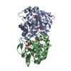
| Title | Crystal structure of Human Orotidine 5'-monophosphate Decarboxylase complexed with BMP | ||||||
 Components Components | Uridine 5'-monophosphate synthase | ||||||
 Keywords Keywords |  LYASE / UMP synthase / C-terminal Domain / Orotidine 5'-monophosphate Decarboxylase / human / BMP / Alternative splicing / Disease mutation / Glycosyltransferase / Multifunctional enzyme / Polymorphism / Pyrimidine biosynthesis / Transferase LYASE / UMP synthase / C-terminal Domain / Orotidine 5'-monophosphate Decarboxylase / human / BMP / Alternative splicing / Disease mutation / Glycosyltransferase / Multifunctional enzyme / Polymorphism / Pyrimidine biosynthesis / Transferase | ||||||
| Function / homology |  Function and homology information Function and homology informationUMP biosynthetic process / orotate phosphoribosyltransferase / orotate phosphoribosyltransferase activity / pyrimidine nucleobase biosynthetic process / Pyrimidine biosynthesis / UDP biosynthetic process / orotidine-5'-phosphate decarboxylase / orotidine-5'-phosphate decarboxylase activity / 'de novo' UMP biosynthetic process / 'de novo' pyrimidine nucleobase biosynthetic process ...UMP biosynthetic process / orotate phosphoribosyltransferase / orotate phosphoribosyltransferase activity / pyrimidine nucleobase biosynthetic process / Pyrimidine biosynthesis / UDP biosynthetic process / orotidine-5'-phosphate decarboxylase / orotidine-5'-phosphate decarboxylase activity / 'de novo' UMP biosynthetic process / 'de novo' pyrimidine nucleobase biosynthetic process / lactation / female pregnancy / cellular response to xenobiotic stimulus / identical protein binding / nucleus / cytosol / cytoplasmSimilarity search - Function | ||||||
| Biological species |  Homo sapiens (human) Homo sapiens (human) | ||||||
| Method | X-RAY DIFFRACTION / SYNCHROTRON / MOLECULAR REPLACEMENT / Resolution: 1.93 Å | ||||||
 Authors Authors | Liu, Y. / Tang, H.L. / Wang, X.Y. / Kotra, L.P. / Pai, E.F. | ||||||
 Citation Citation | Journal: To be Published Title: Crystal structure of Human Orotidine 5'-monophosphate Decarboxylase complexed with BMP Authors: Liu, Y. / Tang, H.L. / Wang, X.Y. / Kotra, L.P. / Pai, E.F. | ||||||
| History |
|
- Structure visualization
| Structure viewer | Molecule: MolmilJmol/JSmol |
|---|
- Downloads & links
-Download
| PDBx/mmCIF format | 3bgg.cif.gz | 67.5 KB | Display | PDBx/mmCIF format |
|---|---|---|---|---|
| PDB format | pdb3bgg.ent.gz | 48.6 KB | Display | PDB format |
| PDBx/mmJSON format | 3bgg.json.gz | Tree view | PDBx/mmJSON format | |
| Others |  Other downloads Other downloads |
-Validation report
| Arichive directory | https://data.pdbj.org/pub/pdb/validation_reports/bg/3bggftp://data.pdbj.org/pub/pdb/validation_reports/bg/3bgg | HTTPS FTP |
|---|
-Related structure data
| Related structure data |  2p1fS S: Starting model for refinement |
|---|---|
| Similar structure data |


-Links
 PDBj
PDBj


- Assembly
| Deposited unit | 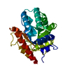
| ||||||||
|---|---|---|---|---|---|---|---|---|---|
| 1 | 
| ||||||||
| Unit cell |
|
-Components
| #1: Protein | Mass: 31751.604 Da / Num. of mol.: 1 / Fragment: Orotidine 5'-phosphate decarboxylase Source method: isolated from a genetically manipulated source Source: (gene. exp.) Homo sapiens (human) / Gene: UMPS / Plasmid: pET15b / Production host:  Escherichia coli (E. coli) / Strain (production host): BL21(DE3)pLysS Escherichia coli (E. coli) / Strain (production host): BL21(DE3)pLysSReferences: UniProt: P11172, orotidine-5'-phosphate decarboxylase |
|---|---|
| #2: Chemical | ChemComp-BMP /   Type: RNA linking / Mass: 340.181 Da / Num. of mol.: 1 / Source method: obtained synthetically / Formula: C9H13N2O10P Type: RNA linking / Mass: 340.181 Da / Num. of mol.: 1 / Source method: obtained synthetically / Formula: C9H13N2O10P |
| #3: Water | ChemComp-HOH / Water Mass: 18.015 Da / Num. of mol.: 132 / Source method: isolated from a natural source / Formula: H2O Mass: 18.015 Da / Num. of mol.: 132 / Source method: isolated from a natural source / Formula: H2O |
-Experimental details
-Experiment
| Experiment | Method: X-RAY DIFFRACTION / Number of used crystals: 1 |
|---|
- Sample preparation
| Crystal | Density Matthews: 2.21 Å3/Da / Density % sol: 44.43 % |
|---|---|
| Crystal grow | Temperature: 293 K / Method: vapor diffusion, hanging drop / pH: 8.4 Details: Ammonium Sulfate, pH 8.4, VAPOR DIFFUSION, HANGING DROP, temperature 293K |
-Data collection
| Diffraction | Mean temperature: 100 K |
|---|---|
| Diffraction source | Source: SYNCHROTRON / Site: APS  / Beamline: 14-BM-C / Wavelength: 0.9002 Å / Beamline: 14-BM-C / Wavelength: 0.9002 Å |
| Detector | Type: ADSC QUANTUM 315 / Detector: CCD / Date: Feb 22, 2007 |
| Radiation | Protocol: SINGLE WAVELENGTH / Monochromatic (M) / Laue (L): M / Scattering type: x-ray |
| Radiation wavelength | Wavelength: 0.9002 Å / Relative weight: 1 |
| Reflection | Resolution: 1.93→50 Å / Num. all: 21306 / Num. obs: 20304 / % possible obs: 95.3 % / Observed criterion σ(F): 0 / Observed criterion σ(I): 0 / Redundancy: 7 % / Rmerge(I) obs: 0.13 / Rsym value: 0.13 / Net I/σ(I): 7.5 |
| Reflection shell | Resolution: 1.93→1.96 Å / Redundancy: 5.6 % / Rmerge(I) obs: 0.5 / Mean I/σ(I) obs: 2.18 / Num. unique all: 818 / Rsym value: 0.5 / % possible all: 74.6 |
- Processing
| Software |
| ||||||||||||||||||||||||||||||||||||||||||||||||||||||||||||||||||||||||||||||||||||||||||
|---|---|---|---|---|---|---|---|---|---|---|---|---|---|---|---|---|---|---|---|---|---|---|---|---|---|---|---|---|---|---|---|---|---|---|---|---|---|---|---|---|---|---|---|---|---|---|---|---|---|---|---|---|---|---|---|---|---|---|---|---|---|---|---|---|---|---|---|---|---|---|---|---|---|---|---|---|---|---|---|---|---|---|---|---|---|---|---|---|---|---|---|
| Refinement | Method to determine structure: MOLECULAR REPLACEMENT Starting model: 2P1F Resolution: 1.93→50 Å / Cor.coef. Fo:Fc: 0.958 / Cor.coef. Fo:Fc free: 0.946 / SU B: 3.976 / SU ML: 0.112 / Cross valid method: THROUGHOUT / σ(F): 0 / σ(I): 0 / ESU R: 0.163 / ESU R Free: 0.141 / Stereochemistry target values: MAXIMUM LIKELIHOOD / Details: HYDROGENS HAVE BEEN ADDED IN THE RIDING POSITIONS
| ||||||||||||||||||||||||||||||||||||||||||||||||||||||||||||||||||||||||||||||||||||||||||
| Solvent computation | Ion probe radii: 0.8 Å / Shrinkage radii: 0.8 Å / VDW probe radii: 1.4 Å / Solvent model: MASK | ||||||||||||||||||||||||||||||||||||||||||||||||||||||||||||||||||||||||||||||||||||||||||
| Displacement parameters | Biso mean: 27.081 Å2
| ||||||||||||||||||||||||||||||||||||||||||||||||||||||||||||||||||||||||||||||||||||||||||
| Refinement step | Cycle: LAST / Resolution: 1.93→50 Å
| ||||||||||||||||||||||||||||||||||||||||||||||||||||||||||||||||||||||||||||||||||||||||||
| Refine LS restraints |
| ||||||||||||||||||||||||||||||||||||||||||||||||||||||||||||||||||||||||||||||||||||||||||
| LS refinement shell | Resolution: 1.93→1.98 Å / Total num. of bins used: 20
|
