 Movie
Movie Controller

[English] 日本語
 Yorodumi
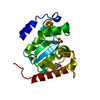
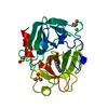
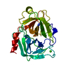
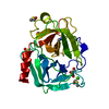
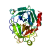
- PDB-3bcn: Crystal structure of a papain-like cysteine protease Ervatamin-A ... -
+ Open data

- Basic information
| Entry | Database: PDB / ID: 3bcn | |||||||||
|---|---|---|---|---|---|---|---|---|---|---|
| Title | Crystal structure of a papain-like cysteine protease Ervatamin-A complexed with irreversible inhibitor E-64 | |||||||||
 Components Components | Ervatamin-A | |||||||||
 Keywords Keywords |  HYDROLASE / protease-inhibitor complex / papain-like fold / plant cysteine protease / Ervatamin / Thiol protease HYDROLASE / protease-inhibitor complex / papain-like fold / plant cysteine protease / Ervatamin / Thiol protease | |||||||||
| Function / homology |  Function and homology information Function and homology informationcysteine-type peptidase activity / Hydrolases; Acting on peptide bonds (peptidases); Cysteine endopeptidasesSimilarity search - Function | |||||||||
| Biological species |  Tabernaemontana divaricata (crepe jasmine) Tabernaemontana divaricata (crepe jasmine) | |||||||||
| Method | X-RAY DIFFRACTION / MOLECULAR REPLACEMENT / Resolution: 2.85 Å | |||||||||
 Authors Authors | Ghosh, R. / Chakrabarti, C. / Dattagupta, J.K. / Biswas, S. | |||||||||
 Citation Citation | Journal: Febs J. / Year: 2008 Title: Structural insights into the substrate specificity and activity of ervatamins, the papain-like cysteine proteases from a tropical plant, Ervatamia coronaria Authors: Ghosh, R. / Chakraborty, S. / Chakrabarti, C. / Dattagupta, J.K. / Biswas, S. | |||||||||
| History |
|
- Structure visualization
| Structure viewer | Molecule: MolmilJmol/JSmol |
|---|
- Downloads & links
-Download
| PDBx/mmCIF format | 3bcn.cif.gz | 85.7 KB | Display | PDBx/mmCIF format |
|---|---|---|---|---|
| PDB format | pdb3bcn.ent.gz | 62.9 KB | Display | PDB format |
| PDBx/mmJSON format | 3bcn.json.gz | Tree view | PDBx/mmJSON format | |
| Others |  Other downloads Other downloads |
-Validation report
| Arichive directory | https://data.pdbj.org/pub/pdb/validation_reports/bc/3bcnftp://data.pdbj.org/pub/pdb/validation_reports/bc/3bcn | HTTPS FTP |
|---|
-Related structure data
| Related structure data | 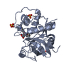 2preC  2pnsS S: Starting model for refinement C: citing same article ( |
|---|---|
| Similar structure data |


-Links
 PDBj
PDBj


- Assembly
| Deposited unit | 
| ||||||||
|---|---|---|---|---|---|---|---|---|---|
| 1 | 
| ||||||||
| 2 | 
| ||||||||
| Unit cell |
|
-Components
| #1: Protein | Mass: 22853.143 Da / Num. of mol.: 2 / Source method: isolated from a natural source Source: (natural) Tabernaemontana divaricata (crepe jasmine)References: UniProt: A5YVK8, Hydrolases; Acting on peptide bonds (peptidases); Cysteine endopeptidases#2: Chemical |   Mass: 360.429 Da / Num. of mol.: 2 / Source method: obtained synthetically / Formula: C15H30N5O5 Mass: 360.429 Da / Num. of mol.: 2 / Source method: obtained synthetically / Formula: C15H30N5O5#3: Chemical | ChemComp-BME / | 2-Mercaptoethanol  Mass: 78.133 Da / Num. of mol.: 1 / Source method: obtained synthetically / Formula: C2H6OS Mass: 78.133 Da / Num. of mol.: 1 / Source method: obtained synthetically / Formula: C2H6OS#4: Water | ChemComp-HOH / | Water Mass: 18.015 Da / Num. of mol.: 40 / Source method: isolated from a natural source / Formula: H2O Mass: 18.015 Da / Num. of mol.: 40 / Source method: isolated from a natural source / Formula: H2OSequence details | THIS PROTEIN IS ISOLATED FROM THE NATIVE SOURCE. THE RESIDUE 12 TO 195 IS FROM CDNA SEQUENCE. CDNA ...THIS PROTEIN IS ISOLATED FROM THE NATIVE SOURCE. THE RESIDUE 12 TO 195 IS FROM CDNA SEQUENCE. CDNA SEQUENCE IS EF591130. 144TH RESIDUE IS GLY FROM THE ELECTRON DENSITY MAP. | |
|---|
-Experimental details
-Experiment
| Experiment | Method: X-RAY DIFFRACTION / Number of used crystals: 1 |
|---|
- Sample preparation
| Crystal | Density Matthews: 2.6 Å3/Da / Density % sol: 52.76 % |
|---|---|
| Crystal grow | Temperature: 293 K / Method: vapor diffusion / pH: 7.5 Details: 0.2M Lithium acetate dihydrate, 20%(w/v) PEG 3350, 12% Glycerol, pH 7.5, VAPOR DIFFUSION, temperature 293K |
-Data collection
| Diffraction | Mean temperature: 100 K |
|---|---|
| Diffraction source | Source: ROTATING ANODE / Type: BRUKER AXS MICROSTAR / Wavelength: 1.5418 Å |
| Detector | Type: MAR scanner 345 mm plate / Detector: IMAGE PLATE / Date: Oct 10, 2007 / Details: Mar multilayer confocal system |
| Radiation | Protocol: SINGLE WAVELENGTH / Monochromatic (M) / Laue (L): M / Scattering type: x-ray |
| Radiation wavelength | Wavelength: 1.5418 Å / Relative weight: 1 |
| Reflection | Resolution: 2.5→100 Å / Num. all: 14392 / Num. obs: 13929 / % possible obs: 85.8 % / Redundancy: 1.97 % / Biso Wilson estimate: 42.7 Å2 / Rmerge(I) obs: 0.0743 / Net I/σ(I): 5.4 |
| Reflection shell | Resolution: 2.5→2.56 Å / Redundancy: 2.04 % / Rmerge(I) obs: 0.1463 / Mean I/σ(I) obs: 3 / Num. unique all: 914 / % possible all: 81.4 |
- Processing
| Software |
| |||||||||||||||||||||||||
|---|---|---|---|---|---|---|---|---|---|---|---|---|---|---|---|---|---|---|---|---|---|---|---|---|---|---|
| Refinement | Method to determine structure: MOLECULAR REPLACEMENT Starting model: 2PNS Resolution: 2.85→30 Å / Data cutoff high absF: 1788723.6 / Data cutoff low absF: 0 / Isotropic thermal model: RESTRAINED / Cross valid method: THROUGHOUT / σ(F): 0 / Stereochemistry target values: Engh & Huber
| |||||||||||||||||||||||||
| Solvent computation | Solvent model: FLAT MODEL / Bsol: 35.0984 Å2 / ksol: 0.356501 e/Å3 | |||||||||||||||||||||||||
| Refine analyze |
| |||||||||||||||||||||||||
| Refinement step | Cycle: LAST / Resolution: 2.85→30 Å
| |||||||||||||||||||||||||
| Refine LS restraints |
| |||||||||||||||||||||||||
| LS refinement shell | Resolution: 2.85→2.98 Å / Total num. of bins used: 8
| |||||||||||||||||||||||||
| Xplor file |
|
